|
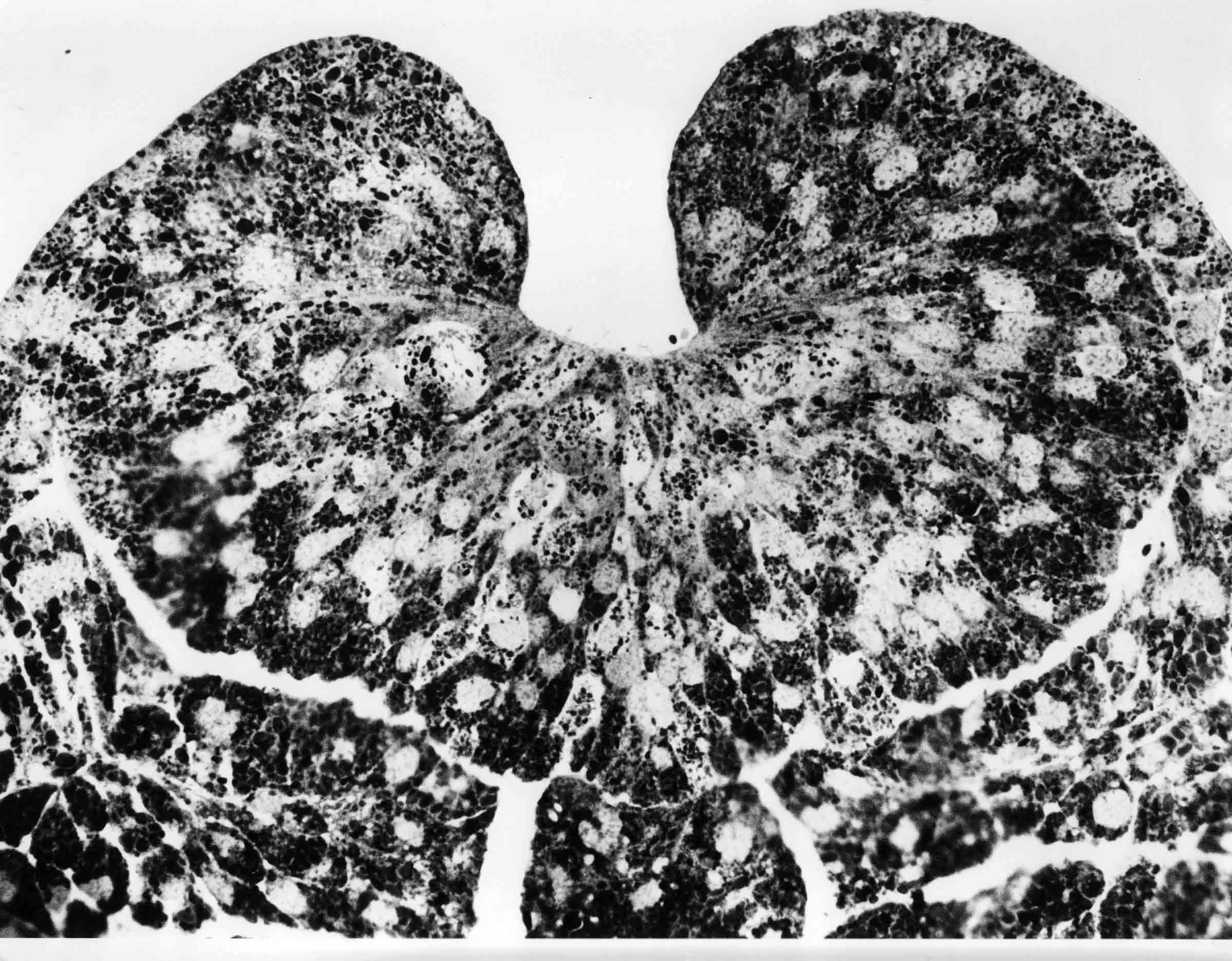
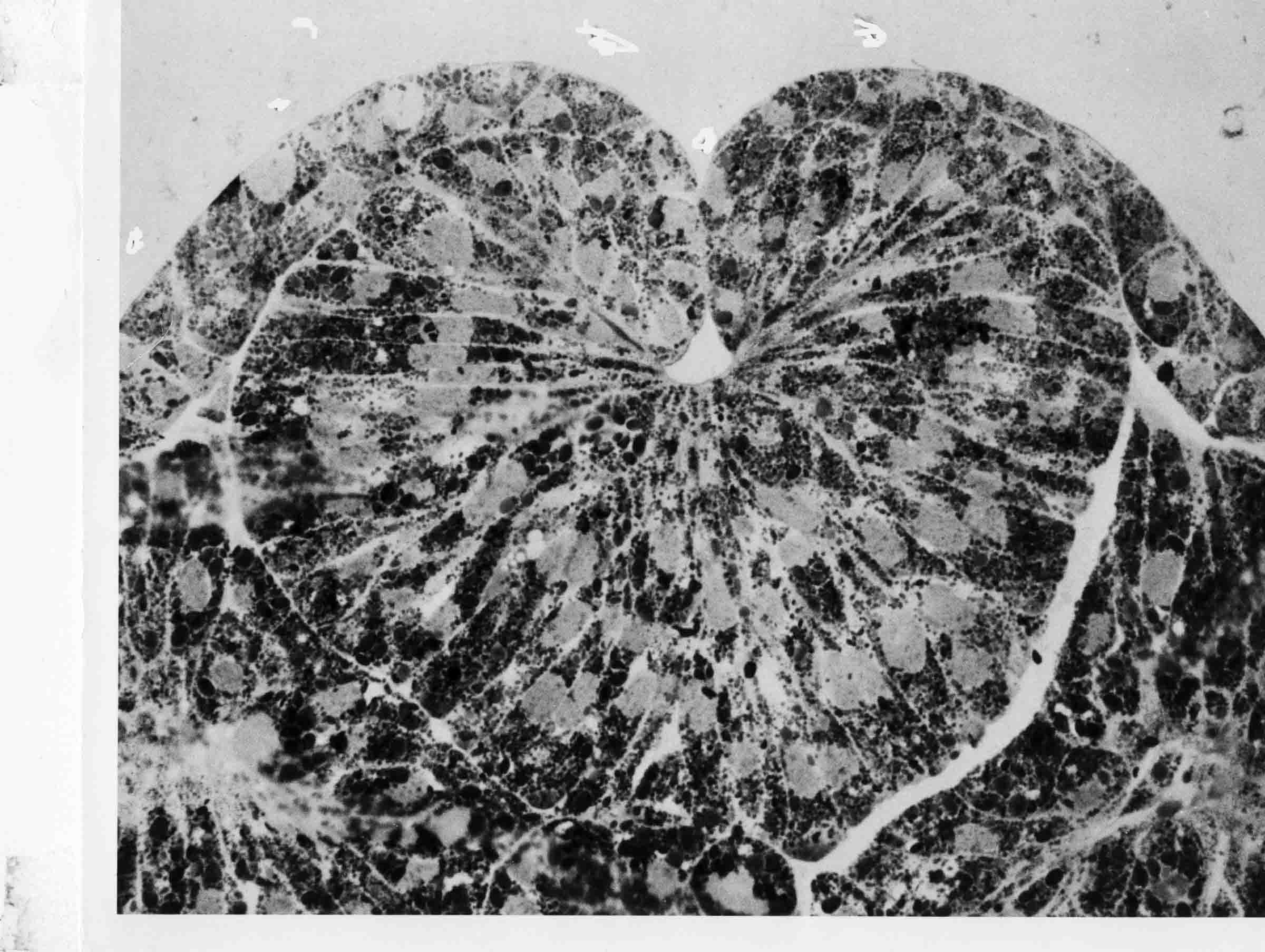
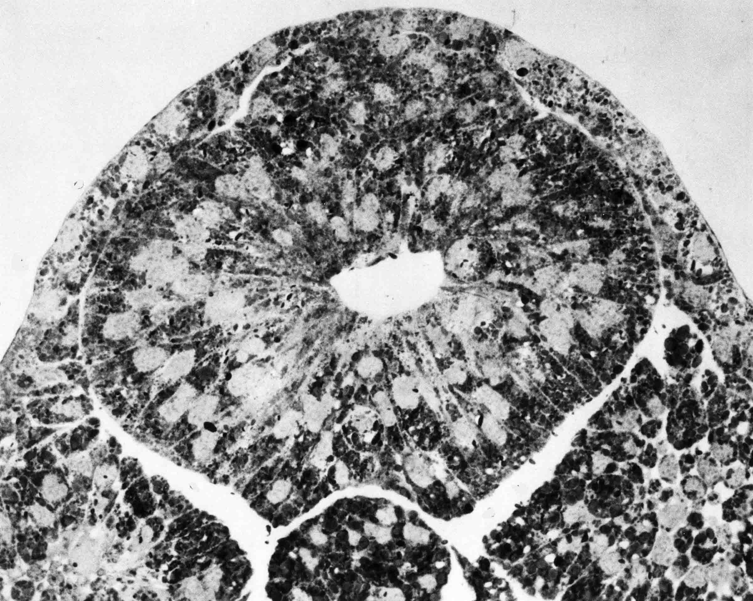
"Thick" Sections Illustrate Neurulation The sections shown in this account are of embryos of Taricha torosa, a West Coast newt. The embryos were fixed and embedded in Epon as for electron microscopy. Sections were cut on an ultramicrotome at a thickness of 2 microns. The sections are called "thick sections". They are actually very thin for cross sections as viewed in ordinary microscopy. The magnifications were about 10X. Each section has a thickness that is within the width of single cells of this material. Cell distribution is thus quite visible. The tractoring of neural plate cells on the epidermis at the neural plate-epidermal boundary is the best explanation of the raising of a neural fold and the rolling of the fold toward the midline of the plate, ending in the closure of the plate into a tube. Many observations and experiments support this assertion. Some of them follow this statement of how tractoring between neural plate cells and epidermis raise the neural folds and then close the folds into a tube. The cells of the neural plate abut cells of the epidermis at the neural plate-epidermis boundary. The two cell types will not mix, and abutting cells are stuck at the boundary by greater adhesion between them, and by contact inhibition. A neural plate cell at the boundary tractors against epidermal cells, elongating the plate cell. The next more-medial plate cell then tractors against the elongating cell and contacts epidermis at its basal end to tractor against the epidermis, and a wave of contraction moves from the basal parts of the cell to the apical end, producing a rolling moment, lifting the epidermis up over the neural plate. This process progresses incrementally mediad. The result is a rising of the epidermis up over the edge of the neural plate producing a neural fold. This rolling moment of the epidermis is quite visible and is not explained by other models. 

This section of a T. torosa neurula shows tractoring and rolling as in the bottom right of the diagram above the sections. 

|