|
In our studies of neurulation, we described how the neural plate cells, at their contact with epidermis, crawl beneath the epidermis, raising a neural fold. I suggested to David Moury that these data imply that a neural fold should arise between any piece of neural plate surgically transplanted against any piece of epidermis. Moury's doctoral research immedietly confirmed this implication. Moury used embryos of the salamander Ambystoma mexicanum, the Mexican axolotl. The publication of this part of the work is: Neural Fold Formation at Newly Created Boundaries between Neural Plate and Epidermis in the Axolotl, J. David Moury and Antone G. Jacobson (1989) Developmental Biology 133:44-57. 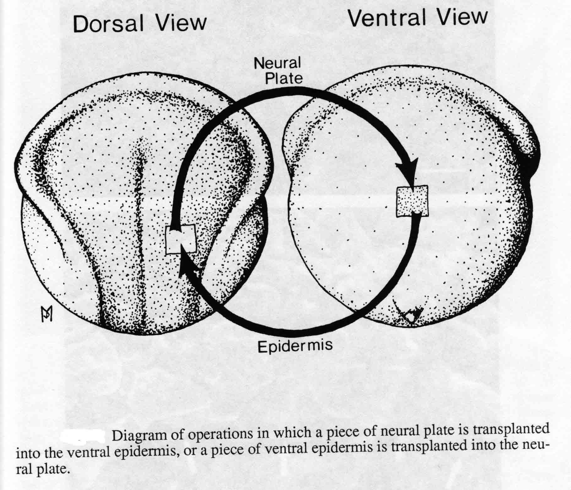 In all cases, the new boundary between neural plate and epidermis formed neural folds surrounding the implant. This is illustrated in the figure below for an implant of neural plate from a normally pigmented embryo into the ventral epidermis of an albino embryo. 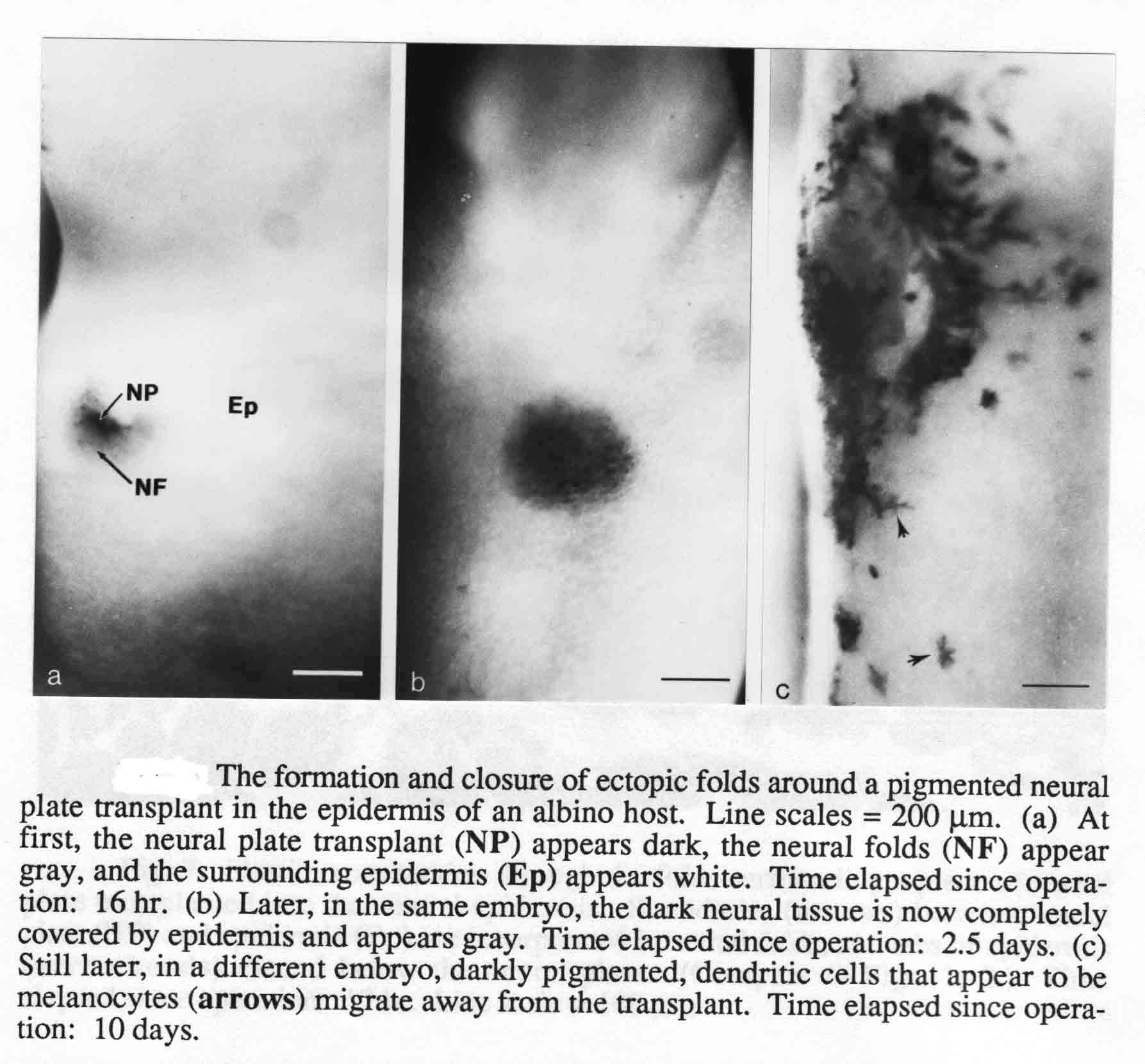 An example of neural folds formed when a piece of epidermis is implanted into a neural plate is shown below. Such epidermal implants always expand rather than contract as surrounding neural plate contracts. 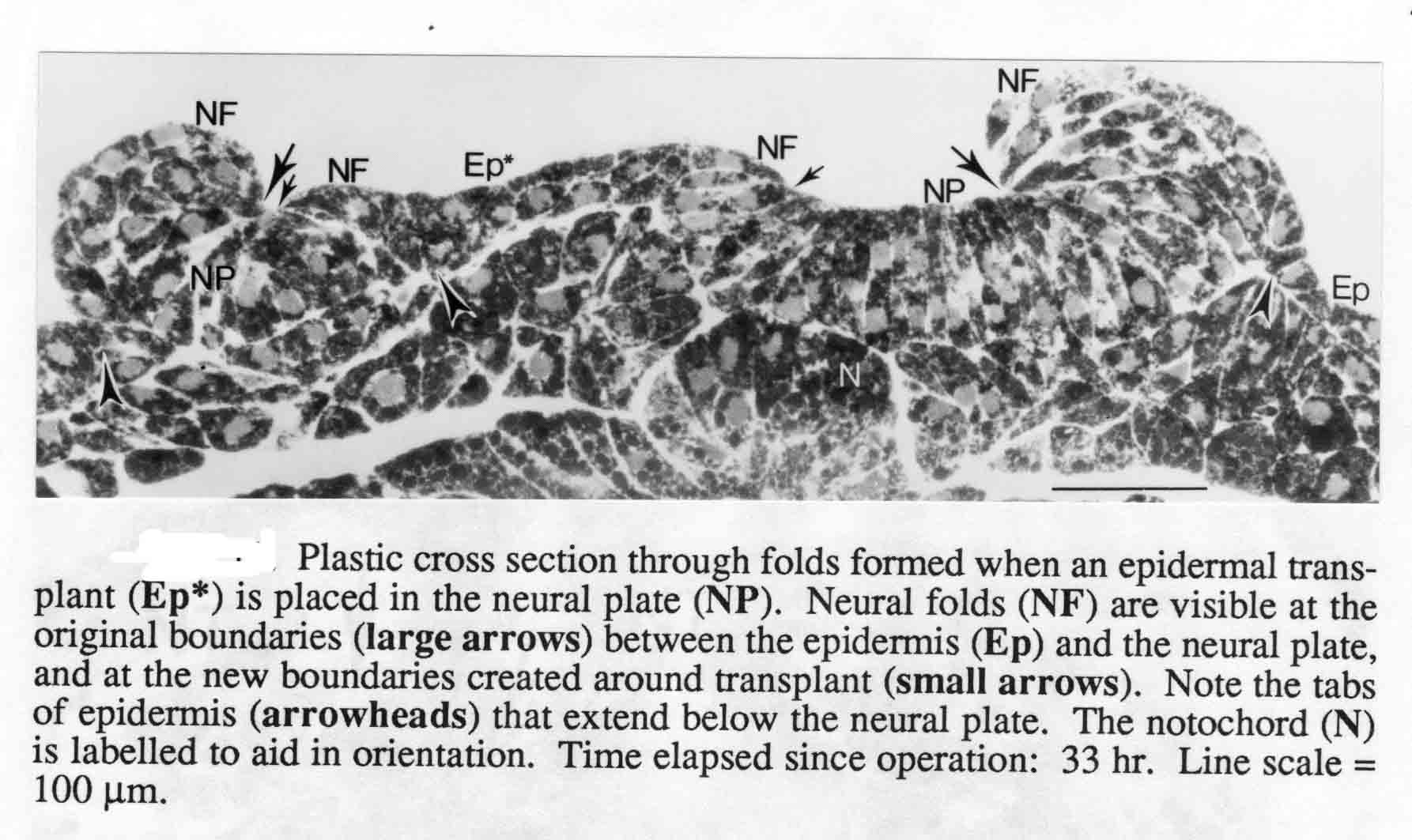 The second part of the Moury study addressed the question of whether neural crest cells originate from the neural plate, the epidermis, or from both of these tissues. Many authors had asked this question with conflicting answers. This part of Moury's work is published at: The Origins of Neural Crest Cells in the Axolotl, J. David Moury and Antone G. Jacobson. (1990) Developmental Biology 141:243-253. Using normally pigmented and albino embryos to distinguish the source of pigmented cells made it possible to discern the source of neural crest cells formed in the new folds. The figure below shows the transplant in place of a piece of the albino host neural fold of pigmented neural plate or epidermis.  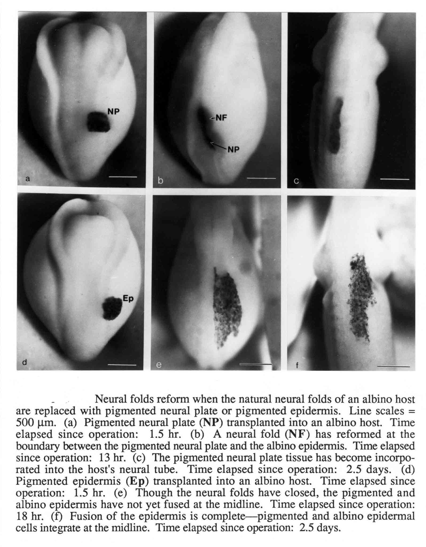 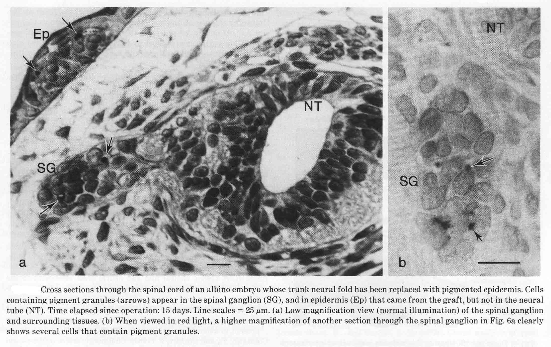 |