 |  |
Glaucous form (Catron Co., NM) | Nonglaucous form (Presidio Co., TX) |
|---|---|
| Fraxinus velutina. Both images at same scale | |
| |
Glaucous form (Catron Co., NM) | Nonglaucous form (Presidio Co., TX) |
|---|---|
| Fraxinus velutina. Both images at same scale | |

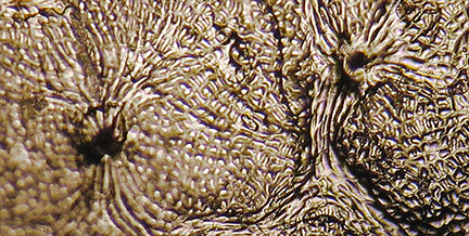
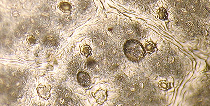
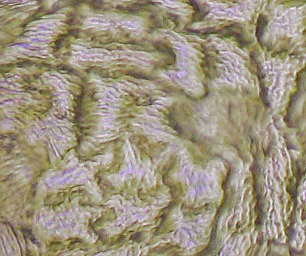
Abaxial leaf surface peels (with clear fingernail polish) were made from numerous Fraxinus leaves, both freshly collected and dried herbarium collections. The peels shown below were photographed at 40X, 100X & 400X with a compound microscope using a handheld Canon Powershot A610 camera and edited in Photoshop — thus images were not always of good quality.
| 40X | 100X | 400X |
|---|---|---|
 |
 |
 |
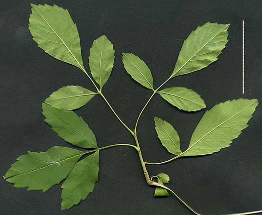
| F. americana (Wisconsin) | ||
 |
 |
 |
| F. albicans (Hays Co., TX) | ||
This network of ridges & papillae also bonded with the nail polish, and considerable effort was required to remove the dried peel for examination, and even then sometimes only small pieces could be obtained. The other ashes in Central Texas do not present this difficulty, and peels sometimes just come loose on their own.
But using leaf peels to distinguish other Fraxinus taxa in Central and West Texas has proved difficult.
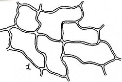
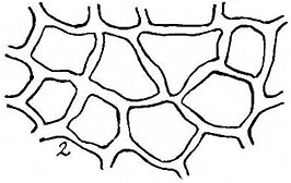
A study by P.A. Munz & J.D. Laudermilk (El Aliso 1949) used epidermal cell shapes to distinguish two western ash taxa:
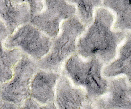
The most significant epidermal character observed by which the two species can be differentiated is the size and shape of the epidermal cells. …F velutina var coriacea, by way of contrast, has very thick–walled cells, the walls being relativel straight so that the cells are quite regular in shape. … data given are for cells away from the veins, since those on or near the veins tend always to be small and rather elongate and regular in shape.
F. oregona F. velutina var. coriacea
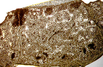
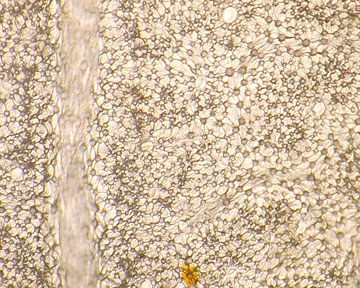
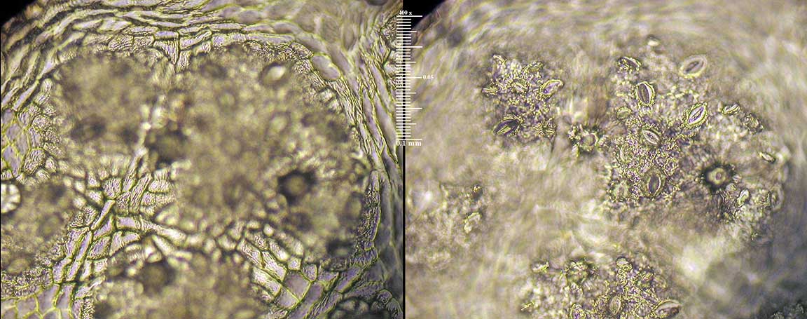
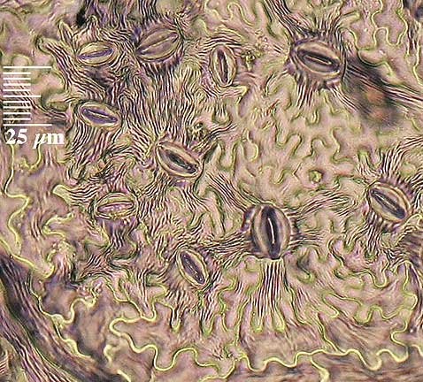
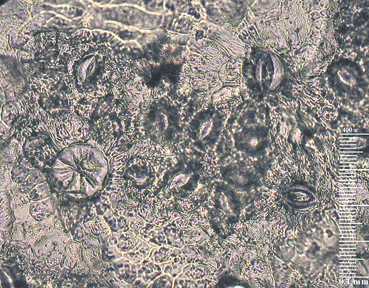
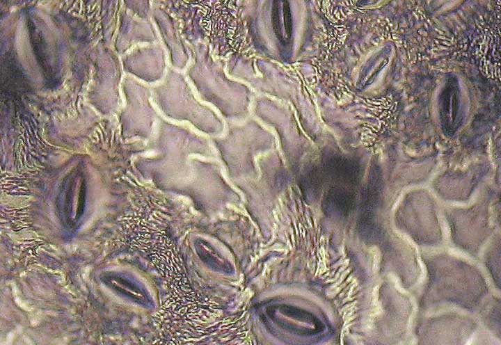
Peels from several collections of F. velutina at TEX show the same pattern indicated by Munz & Laudermilk (100X — 400X):
 |
 |
|---|---|
| Presidio Co., TX (100X) | Catron Co., NM (400X) |
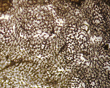
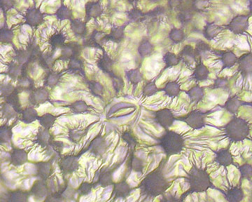
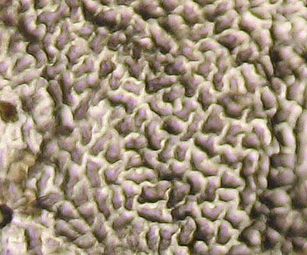
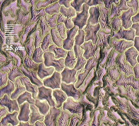
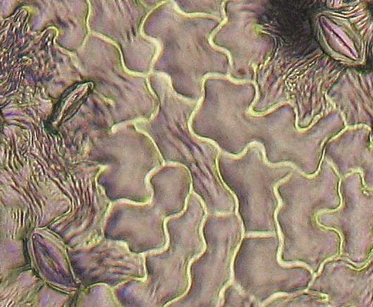
In contrast stands a peel from a specimen currently determined as F. velutina. It's abaxial leaflet surface seems to be covered with papillae. The cell walls lack a uniform shape and are not quite as thick as those above. (100X — 400X)
 |  | |
|---|---|---|
| F. papillosa?, Coconino Co., AZ | ||
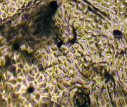
Similarly, the following collection from Presidio Co., TX, lacks the features of the above Presidio F. velutina collection. (100X — 400X)
 |  |
|---|
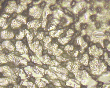
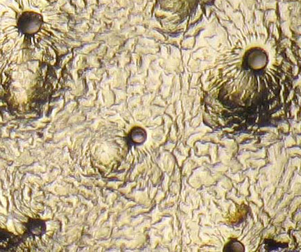
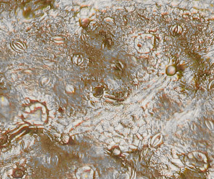
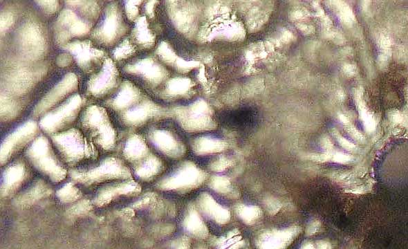
In all 17 collections at TEX/LL, previously determined as F. velutina, were examined. Initially it seemed that two distinct forms were found: (1) heavy glaucous surface with stomata few and far between and very few scales (6 trees); (2) surface not clearly glaucous, cuticle covered with fine striations (except on veins), numerous stomata and scales (11 trees). But subsequently, with a F. berlandieriana tree, I found both types represented. Although the cell structure is clearest with the strong glaucous abaxial surface, the forms with numerous stomata visible, also permit some idea of the cell form — although these cells tend to be close to veins, and may not be typical.
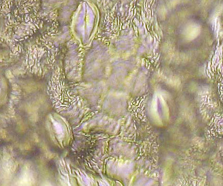
Two images of the same section of F. velutina taken at 400X with two different focal points. It is of the nonglaucous type from Jeff Davis, Co.:
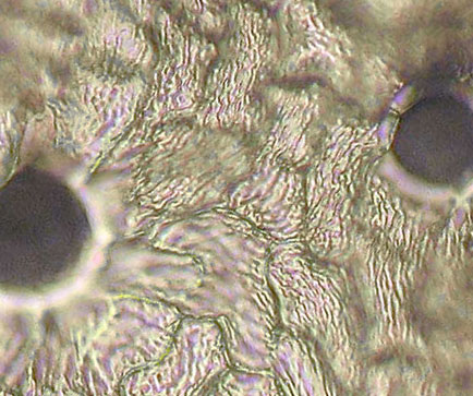
This non-glaucous collection from Brewster Co., TX, also lacks the F. velutina cell features noted above (100X — 400X):
 |  |
|---|
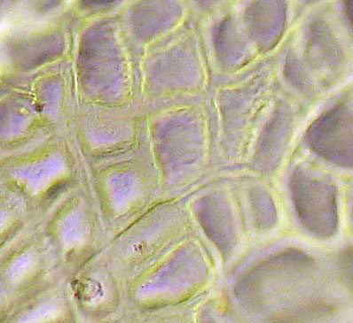
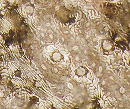
But this collection from Presidio Co. seems to have the expected F. velutina cell form:
 |  |
|---|
As does this collection from Chihuahua, Mexico:
Although most F. berlandieriana leaflets did not show a strongly glaucous surface similar to F. velutina, one such leaflet was found on the same plant with other leaves having nonglaucous features. Both leaflets had strongly striated cuticle surfaces. For herbarium specimens examined this dense striated cuticle surface generally prevented examination of cell forms not along a vein.
 |  |
|---|
Collection from Cameron Co.:
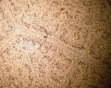
F. pennsylvanica is unlike any of the above, with irregular shaped relatively thick–walled and rounded cells. It does not seem to have a glaucous abaxial form. The following is from the bank of Barton Creek in Austin, TX:
(1) A small ash of unknown origin on the U. Texas campus has leaves with coarsely serrate distal margins, and numerous 3–leaflet leaves, and also has deeply sinused abaxial cells. I am tempted to identify it as F. berlandieriana.
 |  |
|---|