In his A Monograph of the Genus Evolvulus (1934) van Ooststroom describes venation for E. sericeus):
This would appear to match E. P. Klucking's ‘intermediate’ venation pattern (The classification of leaf venation patterns, 1995 (p. 39):
The Intermediate system consists mostly of venation which is a combination of apically directed palmate secondary veins and laterally directed pinnate ones. The apically directed secondaries occur in the lower part of the leaf and the pinnate veins in the upper part.In working with pressed herbarium specimens determining venation was relatively difficult for several reasons. First, the narrower leaves of those forms with glabrous surfaces generally fold when collected, and unless care had been taken prior to drying, the glabrous upper surface is mostly not visible. A number of collections had no visible upper surface or only a small corner of one. Note how this natural folding occurs in the freshly collected stem 'A' in the image below.
Second, with fresh leaves the upper-surface vein impression of the lateral veins was often quite faint, apparent only by virtue of slight color differences. It probably would not maintain its visibility with drying and aging of the leaf surface.
Finally, for unknown reasons many, if not most, leaves that did not fold had been mounted face down.
Consequently I made a short study with fresh stems collected 26 September 2012 from N. Hays Co., from two somewhat different environments: A, from a xeric environment with shallow soil; B, from a grassy area with deep soil. The two stems are shown below with leaves enumerated from bottom to top. Each leaf was forced flat and inspected for visible venation with a dissecting microscope. A number of different types were noted and images were made of each type. The results and images are presented below. [For a survey of 99 leaves, including the two shown here.]
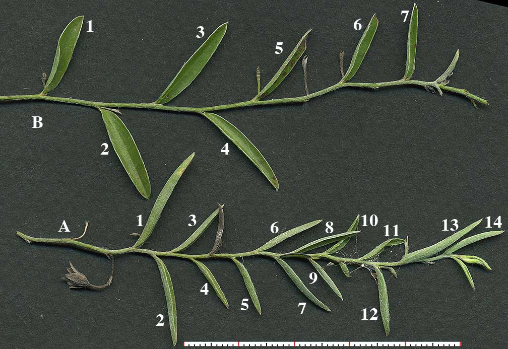
Click on image for extended view.
Thumbnails are full size.
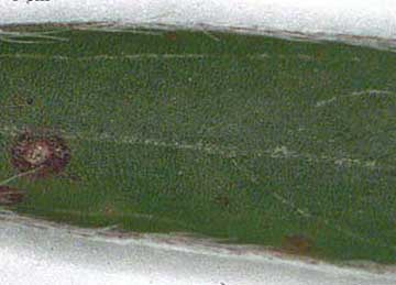
A:leaf no. 1 Basal veins to mid leaf; several faint pinnate veins above the base. |
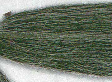
A:leaf no. 1 The scope seemed to show lateral veins, but the image doesn't make it clear. |
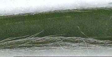
A:leaf no. 2 Faint basal veins. |
|---|
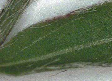
A:leaf no. 4 Faint basal vein on one side only. |
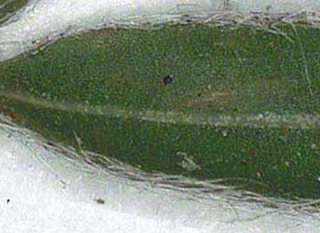
A:leaf no. 6 No lateral vein visible. |
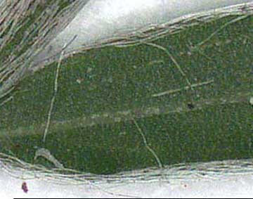
A:leaf no. 8 Faint basal vein on one side only. |
|---|
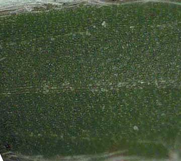
A:leaf no. 9 Faint basal venation. |
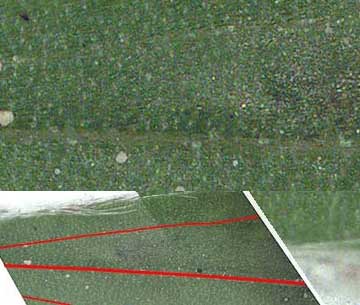
A:leaf no. 13 'Ghost' (barely visible?) basal venation |
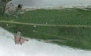
A:leaf no. 14 Perfect basal venation. |
|---|
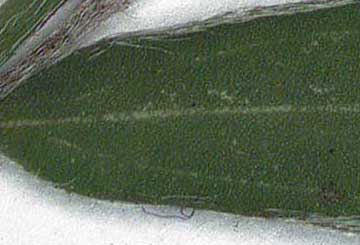
B:leaf no. 2 Basal venation |
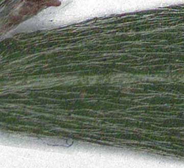
B:leaf no. 2 Abaxial basal venation visible. |
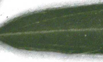
B:leaf no. 4 Basal venation. |
|---|