Vessel to Ray Transport
by Bob Harms (

)
The images below are presented to illustrate the water transport from root to rupture at the stem cortex in our two species. Although several images are from other species, the secondary xylem anatomy is assumed to be essentially the same. I am indebted to Jim Henrickson of the Plant Resources Center for assistance in identifying the structures presented below.
-
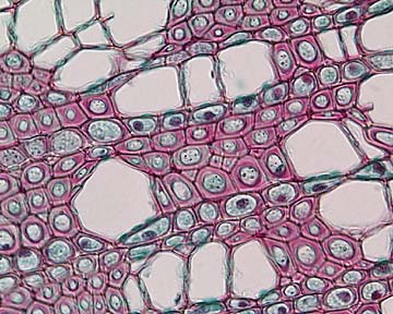
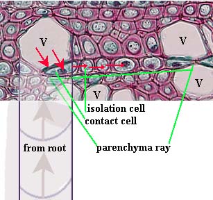
The image on the left is captioned "Sambucus xylem in transverse section" [Paul Schulte, Atlas of Plant Anatomy, Sambucus is 'elderberry']. On the right I have interpreted it as having ray parenchyma as labeled on the right, with imagined flow from a vessel, carrying water from the roots, to a contact cell. Other cells in the section slice probably consist of axial parenchyma and fibers.
-
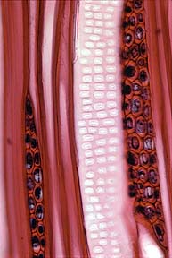
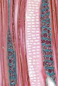
The longitudinal section on the left has the caption "Liriodendron vessel element, showing elongate bordered pits." [BOTANICAL SOCIETY OF AMERICA'S ONLINE IMAGE COLLECTION, Liriodendron is 'American tulip tree'] The cells I have colored blue are axial rays.
-
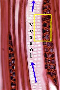
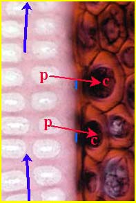
In the above images I have interpreted the area in yellow as flow from pits into contact cells. The pits shown are bordered pits and the pits in contact with contact ray cells are not shown. I assume that pits (P) are aligned in contact with contact ray cells (C) at the blue lines.
-
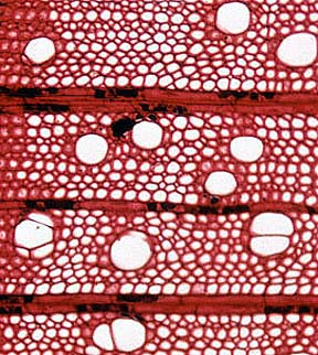
The image on the left is captioned "Acer wood (secondary xylem)". [BOTANICAL SOCIETY OF AMERICA'S ONLINE IMAGE COLLECTION, Acer is 'maple'] The image on the right provides a hypothetical vessel to ray transfers (red arrows) at two points. The smaller vessel cells in cross section result from the cut having been made closer to their endpoints.
-
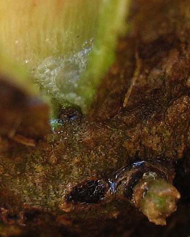
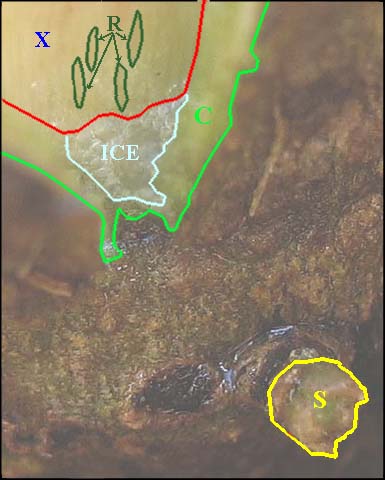
The ruptured base of Verbesina virginica upon melting after a frost event is shown below with an interpretation on the right. X = secondary xylem, R = rays at point of sap egress, C = cortex, S = emerging new shoot.
-
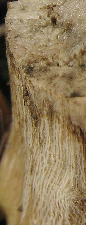
The exposed secondary xylem of Pluchea odorata following detachment of the cortex (etc.). Axial rays can be seen on the longitudinal surface (compare the above image from V. virginica ).