|
West Coast Newt, Taricha torosa The mesonephrogenic kidney mesoderm of the newt lies in the mesoderm mantle between the prospective somites above and the lateral plate mesoderm below, as shown in the map below from his paper. 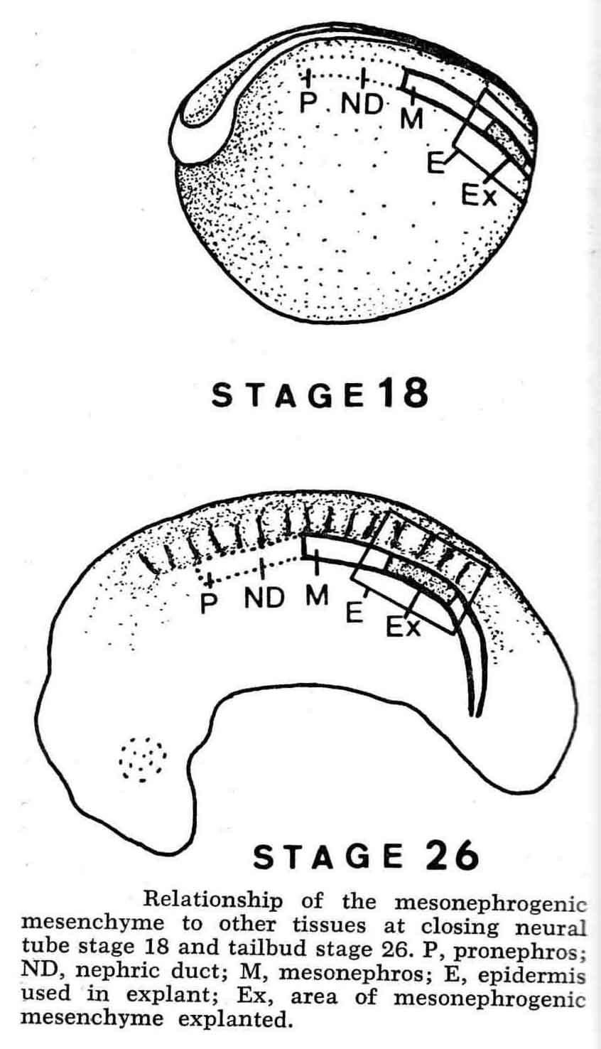 Explants of the mesonephrogenic mesemchyme were made in overlying epidermis, and with or without putative inductor or suppressor tissues. He found the underlying endoderm to be the primary inductor tissue, and the neural crest to be a powerful suppressor tissue. Positive cases were identified by the presence of kidney tubules in sections. Many tissues were examined for their inductive or suppressive effects The nephric duct is a powerful inductor of the kidney, but many explants were sufficiently induced to form kidney tubules long before appearance of the duct. The cumulative effects of several inductor and suppressor tissues control the elicitation of kidney tubules. An interesting case is the following comparison. When mesonephric mesenchyme is explanted in overlying epidermis at stage 22, 27% of the cases formed kidney tuules. When the entire neural fold, and thus all the neural crest cells are removed at stage 16, then the mesonephric mesenchyme of these crest-free embryos was explanted at stage 22, 78% of the cases formed kidney tubules. The complexities of these several tissues influencing the mesonephric mesoderm in additive ways is shown in the graph below: 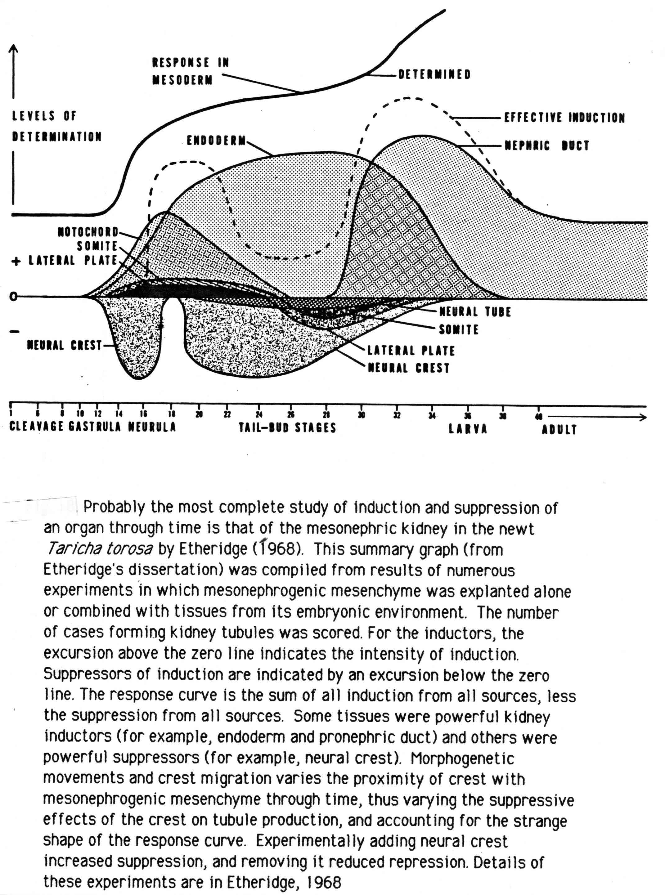 |