|
From over 1,000 sem's, examples have been chosen to help illustrate the development of the newt Taricha torosa. The Twitty-Bodenstein normal staged series for Taricha torosa are shown below. 
Stage 1 is the fertilized egg, the zygote, which is a single cell about 2.4 mm in diameter. Stages 2 through 5 and beyond are cleavage stages, during which simultaneous cell divisions occur. Each division reduces the volume of each dividing cell by half. Each daughter cell inherits an aliquot of yolk platelets so cells or groups of cells can be cultured with internal yolk resources providing nourishiment. There is no growth during the stages shown. Beginning at stage 5, an internal fluid-filled blastocoel cavity forms so the embryo is now a blastula stage as well as a cleavage stage. Blastula stages continue to stage 9 when gastrulation begins and gastrula stages continue to stage 13 when neurula stages begin which last to stage 20. Stages 21 to 40 are early larval stages when the major organ systems are being established. The heart, the first organ to become functional, first beats at stage 34. The embryo hatches and becomes free living at stage 40. Our collection of sem's begins at stage 9; the beginning of gastrulation. To see a video of cleavage stages in the newt, go to Cleavage (six minutes). The entire development of a related salamander is at Development of a Salamander (8.5 minutes). The sem's are each described in the legends beneath the pictures. 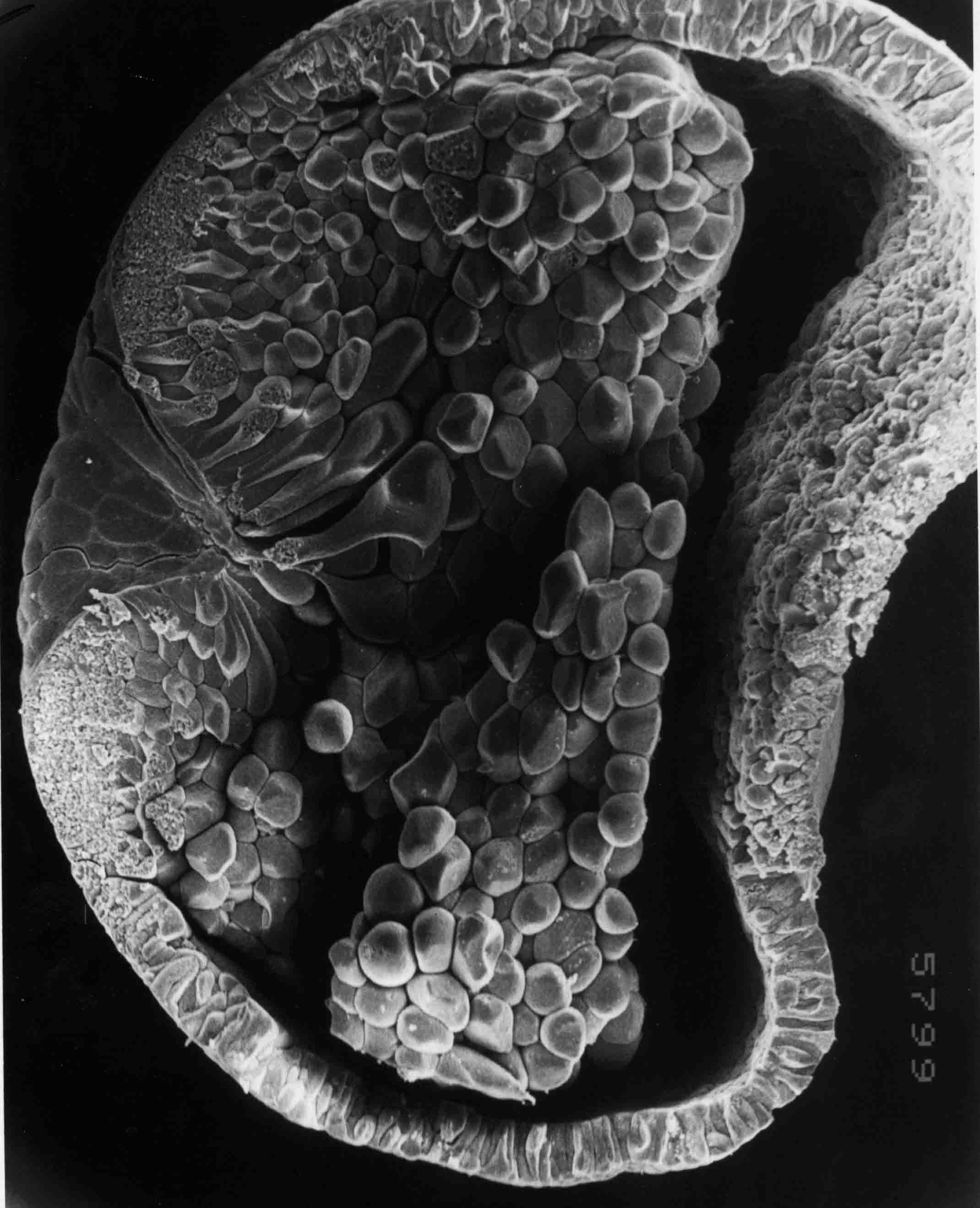 This early stage 9 embryo was prepared for sem, then cut in two and the left half is shown in interior
view. Dorsal is upward, and posterior is to the left where the beginning of the dorsal lip of the
blastopore is forming. The outer surface of the blastula has been pulled inward by the bottle cells
whose apical ends are part of the surface of the embryo. The basal ends of the bottle cells are
migrating toward the anterior end of the embryo where they will become the most-anterior endoderm.
The anterior side (right) has collapsed into the bastocoel cavity in this dissected embryo. The normal
embryo is spherical at this stage. Magnification is 59X
This early stage 9 embryo was prepared for sem, then cut in two and the left half is shown in interior
view. Dorsal is upward, and posterior is to the left where the beginning of the dorsal lip of the
blastopore is forming. The outer surface of the blastula has been pulled inward by the bottle cells
whose apical ends are part of the surface of the embryo. The basal ends of the bottle cells are
migrating toward the anterior end of the embryo where they will become the most-anterior endoderm.
The anterior side (right) has collapsed into the bastocoel cavity in this dissected embryo. The normal
embryo is spherical at this stage. Magnification is 59X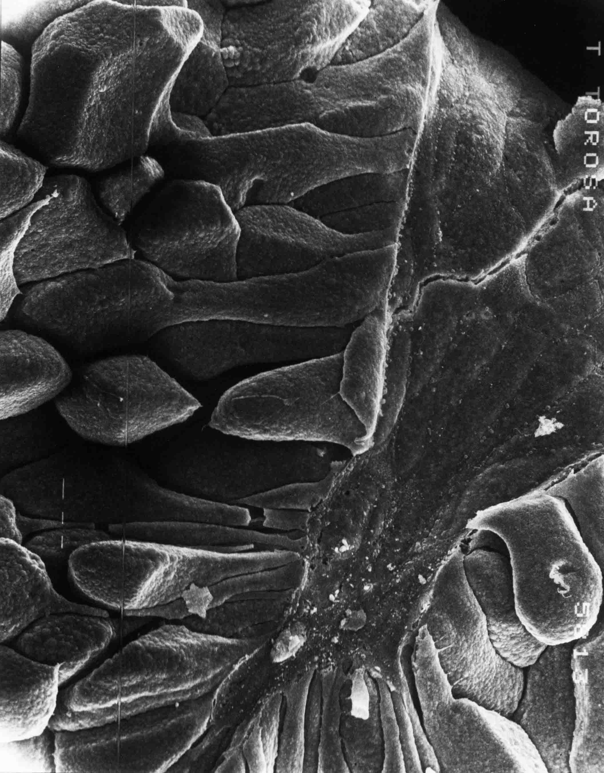
 Higher magnifcation (420 X) of apical ends of bottle cells that have broken away from the surface of
the embryo.
Higher magnifcation (420 X) of apical ends of bottle cells that have broken away from the surface of
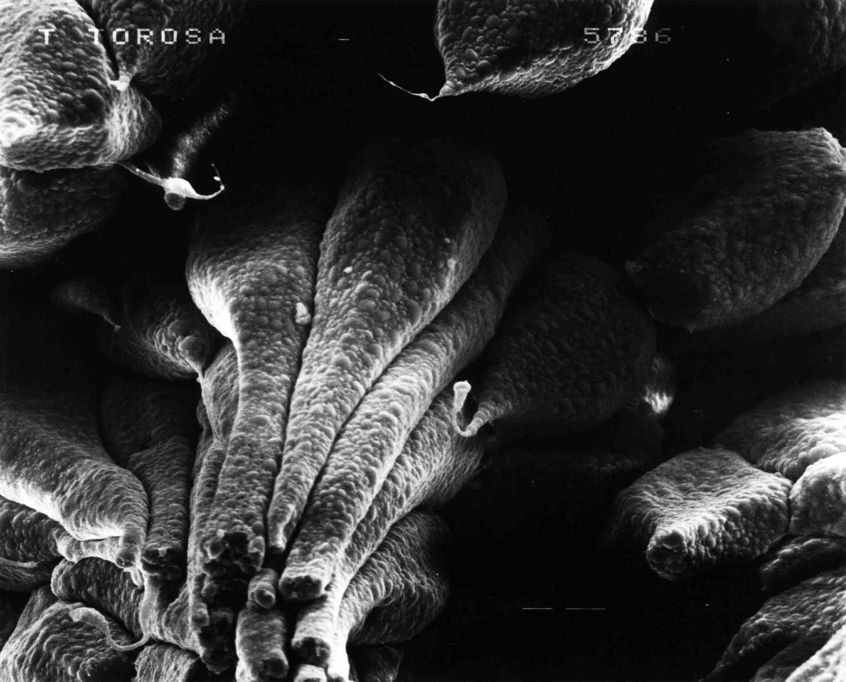
the embryo.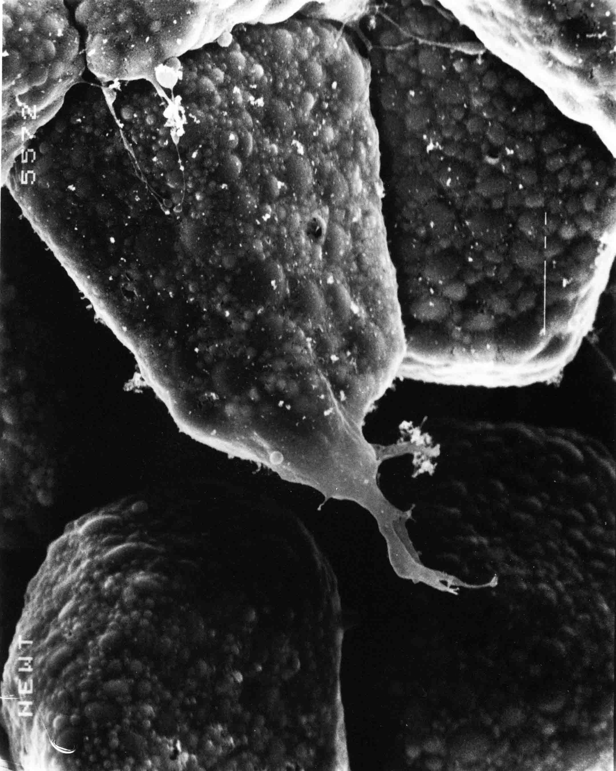 Stage 9; 1150X. A cell process protrudes from the basal end of a bottle cell. The blunt basal end
of a migrating bottle cell is the leading edge.
Stage 9; 1150X. A cell process protrudes from the basal end of a bottle cell. The blunt basal end
of a migrating bottle cell is the leading edge.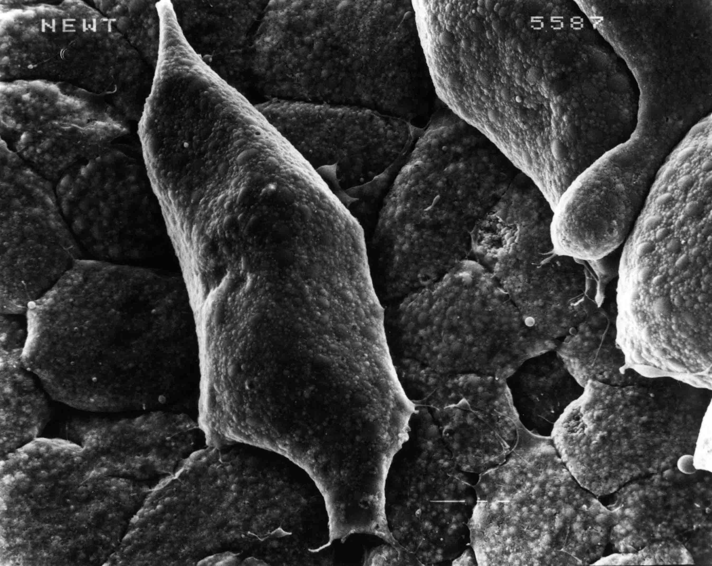 Stage 10; 576X. This bottle cell, now loose from the surface, is migrating against the outer layer
of cells of the embryo. The leading basal end of the cell has protrusions, and there are also small
protrusions from the lateral edges of the cell. The former apical end of the cell is the trailing
edge.
Stage 10; 576X. This bottle cell, now loose from the surface, is migrating against the outer layer
of cells of the embryo. The leading basal end of the cell has protrusions, and there are also small
protrusions from the lateral edges of the cell. The former apical end of the cell is the trailing
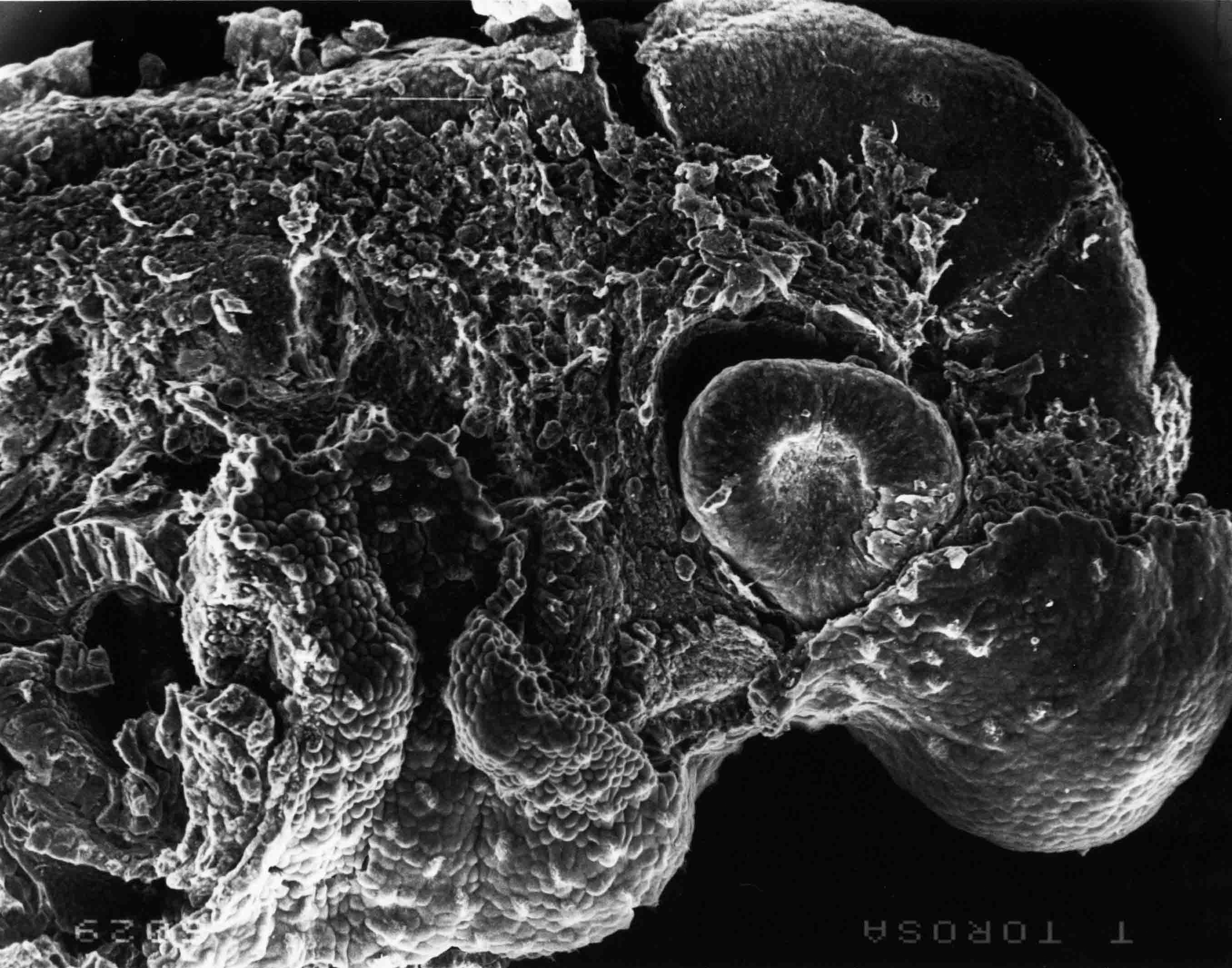
edge.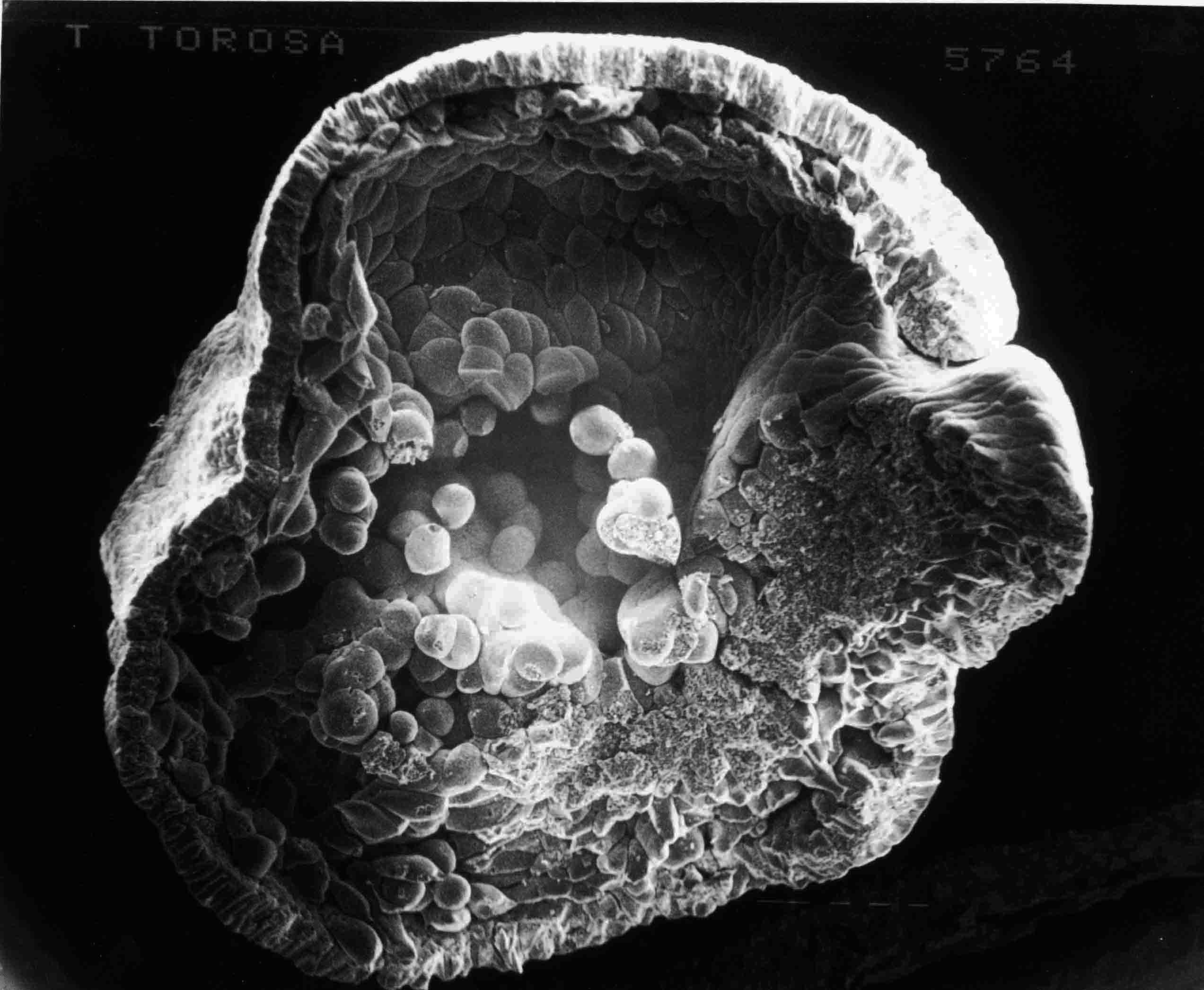 This is a sagittal section of a stage 10+ embryo (40X). One sees the inside of the right half of
the embryo, dorsal lip of the blastopore to the right, dorsal at top, The bottle cells have now
migrated well toward the anteror end of the embryo.
This is a sagittal section of a stage 10+ embryo (40X). One sees the inside of the right half of
the embryo, dorsal lip of the blastopore to the right, dorsal at top, The bottle cells have now
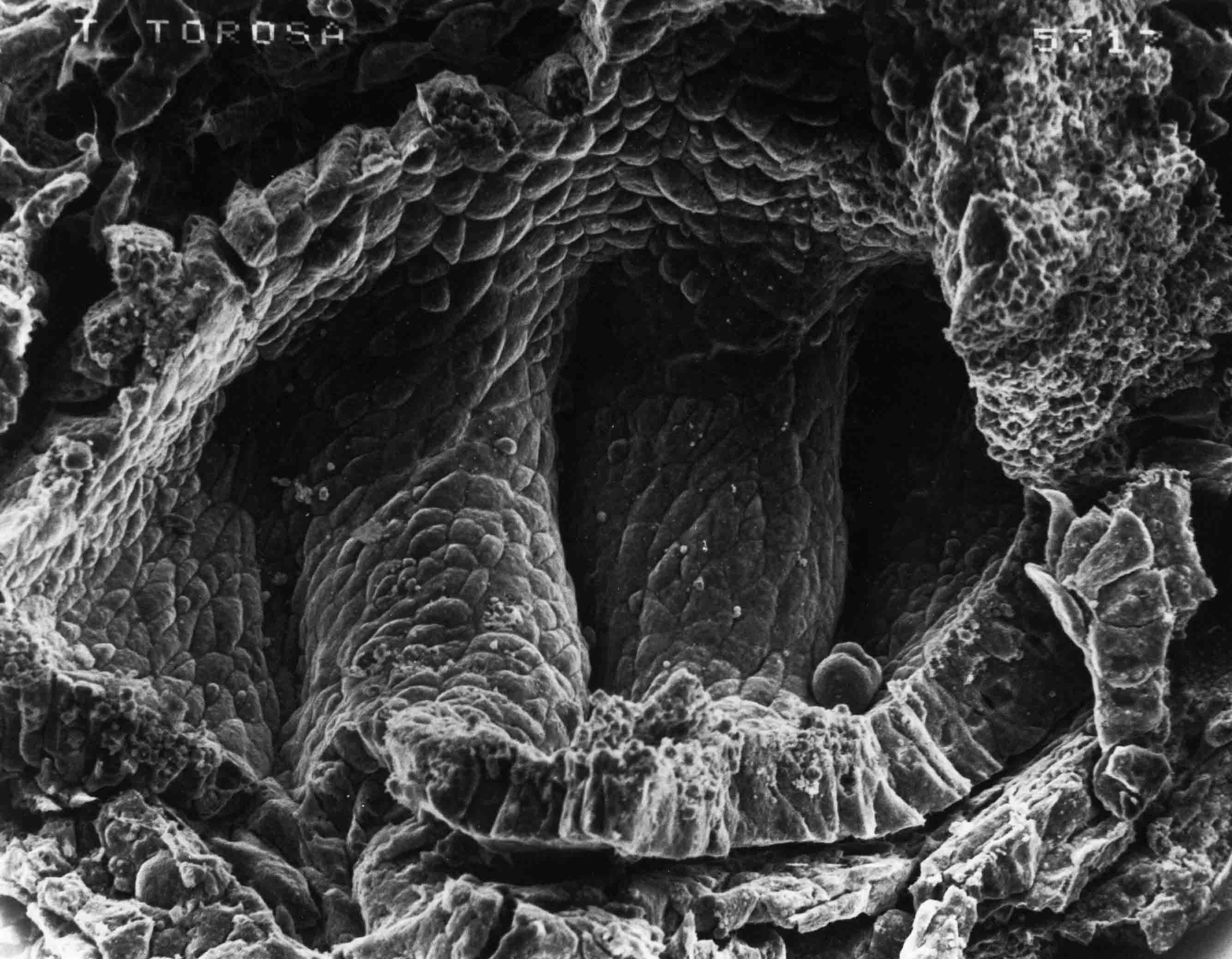
migrated well toward the anteror end of the embryo.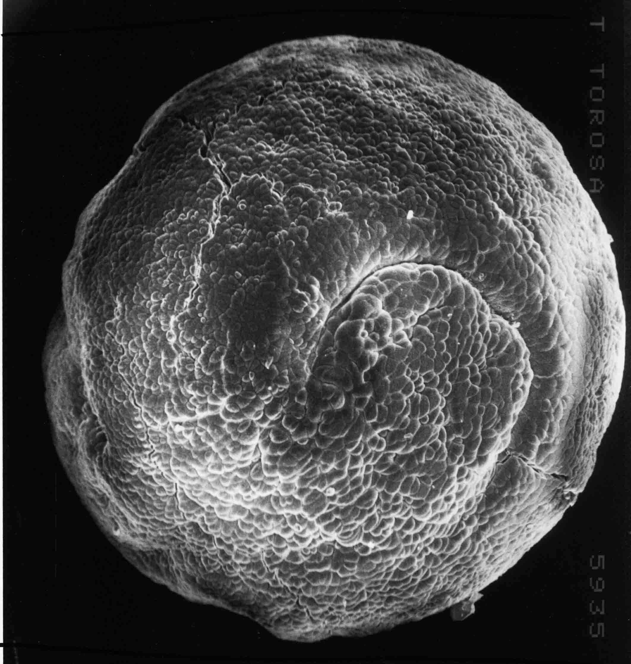 Stage 11+; 42X. This is a posterior view of a stage 11+ embryo. The dorsal lip of the blastopore
continues laterally as the lateral lips of the blastopore. The future neural plate is dorsal to the
dorsal lip, but also extends laterally to each side of the lateral lips. Directly above and to the
sides of the lips are mesodermal cells still involuting around the lips. Endodermal cells are
involuting beneath the lips.
Stage 11+; 42X. This is a posterior view of a stage 11+ embryo. The dorsal lip of the blastopore
continues laterally as the lateral lips of the blastopore. The future neural plate is dorsal to the
dorsal lip, but also extends laterally to each side of the lateral lips. Directly above and to the
sides of the lips are mesodermal cells still involuting around the lips. Endodermal cells are
involuting beneath the lips. 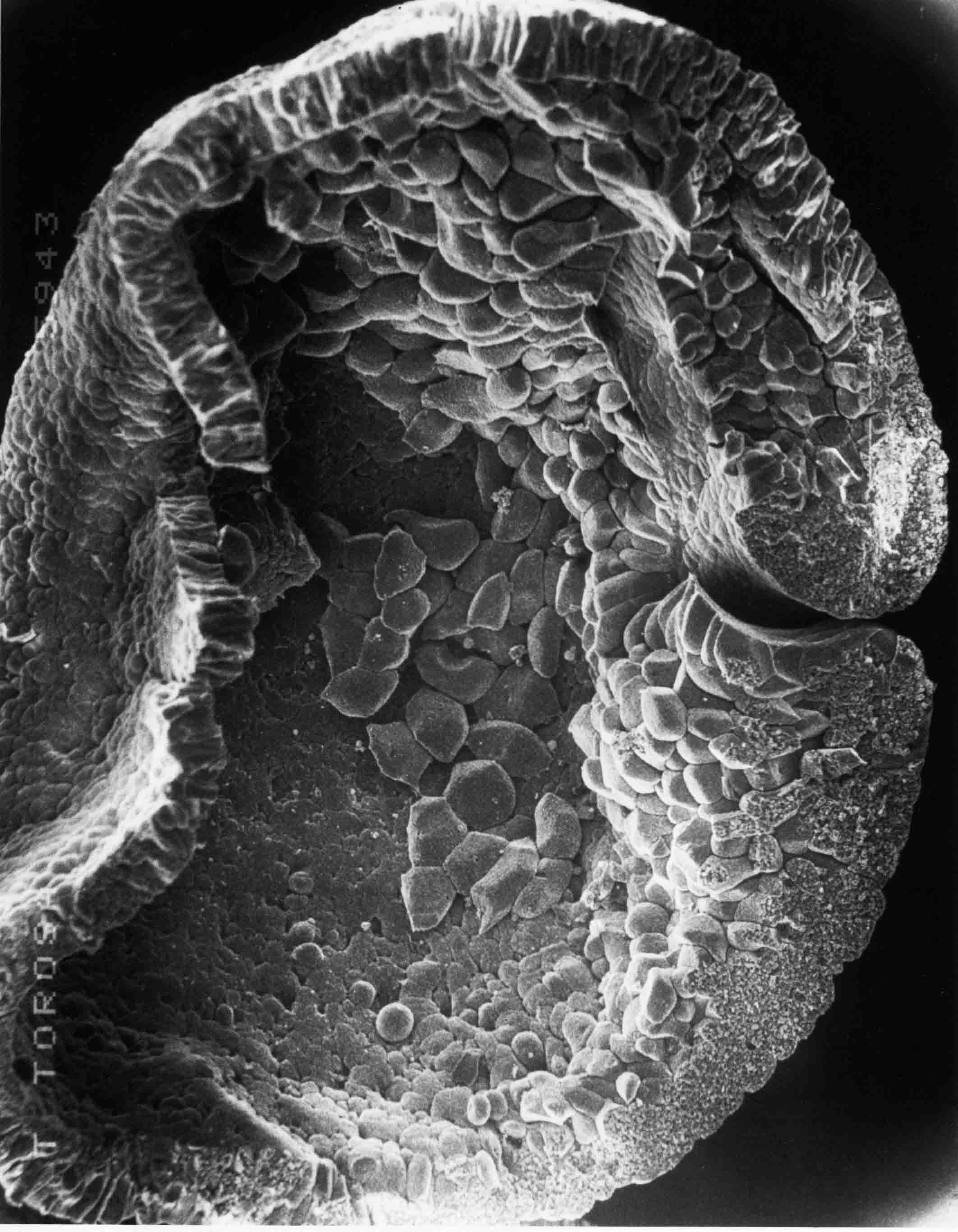 Sagittal section of a stage 11 embryo (59X). One sees the inside of the right half of the embryo.
The embryo now has lateral lips as well as a dorsal lip to the blastopore. Some of the former outside
surface of the embryo now extends far inside the embryo.
Sagittal section of a stage 11 embryo (59X). One sees the inside of the right half of the embryo.
The embryo now has lateral lips as well as a dorsal lip to the blastopore. Some of the former outside
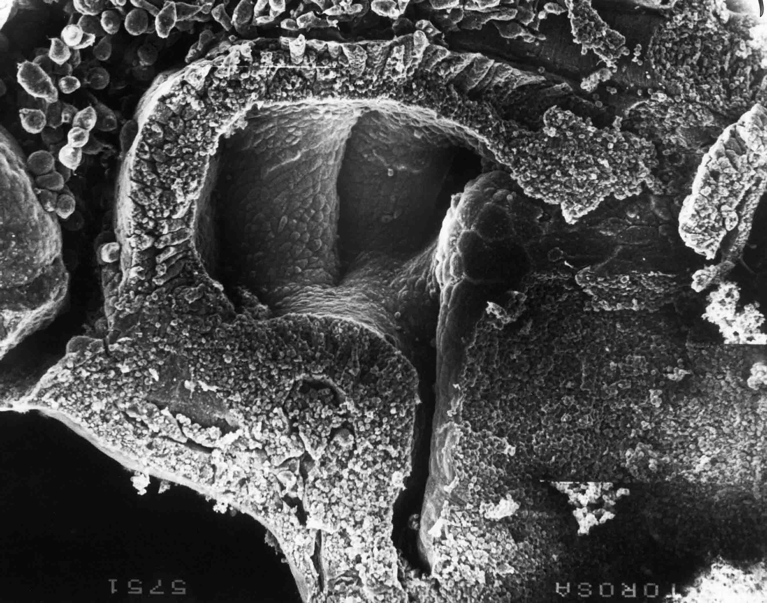
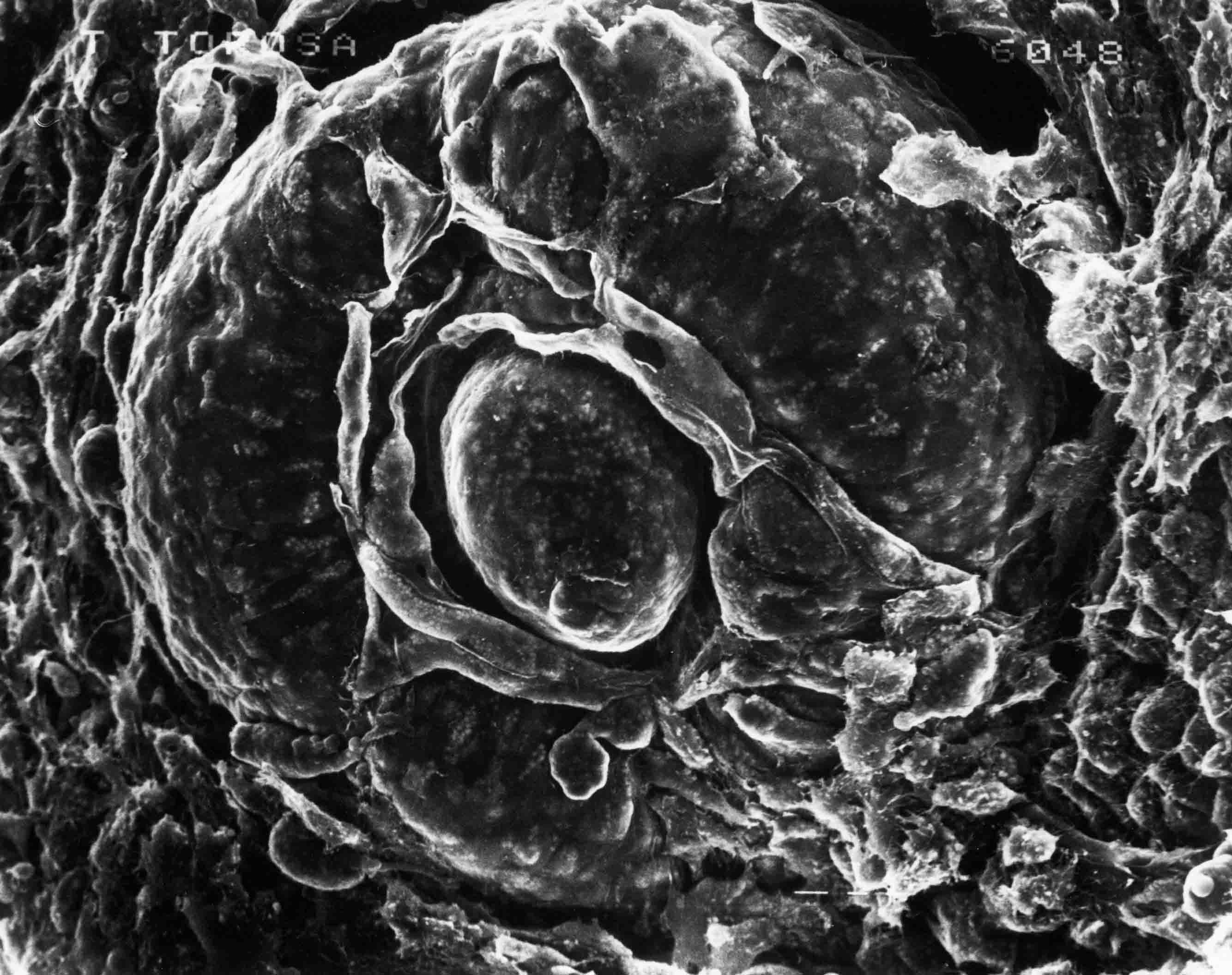
surface of the embryo now extends far inside the embryo.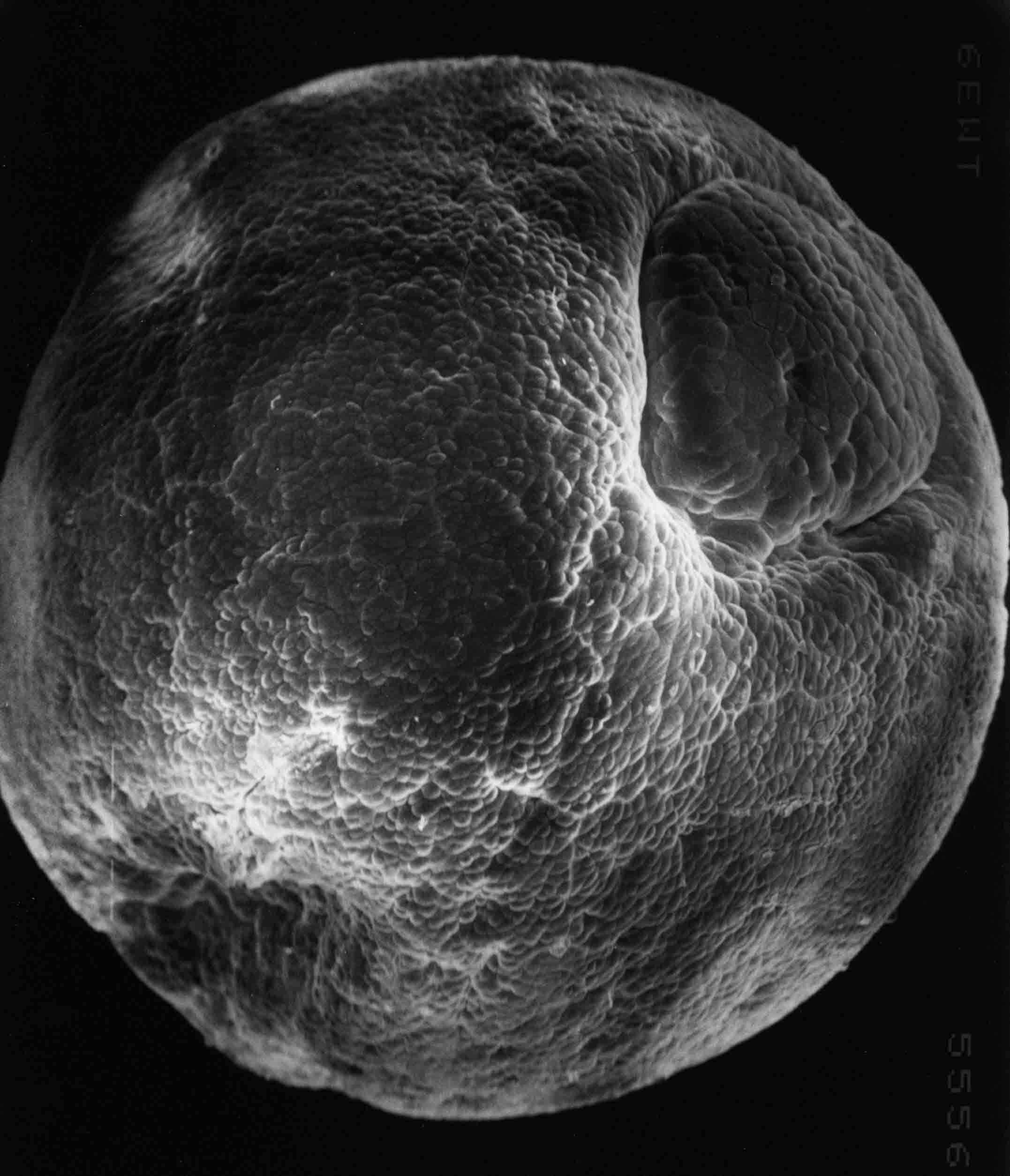 Stage 12; 50X. This is a surface view of the left-posterior side. The ventral lip has formed
completing the circular blastopore which encompasses the large yolk plug.
Stage 12; 50X. This is a surface view of the left-posterior side. The ventral lip has formed
completing the circular blastopore which encompasses the large yolk plug.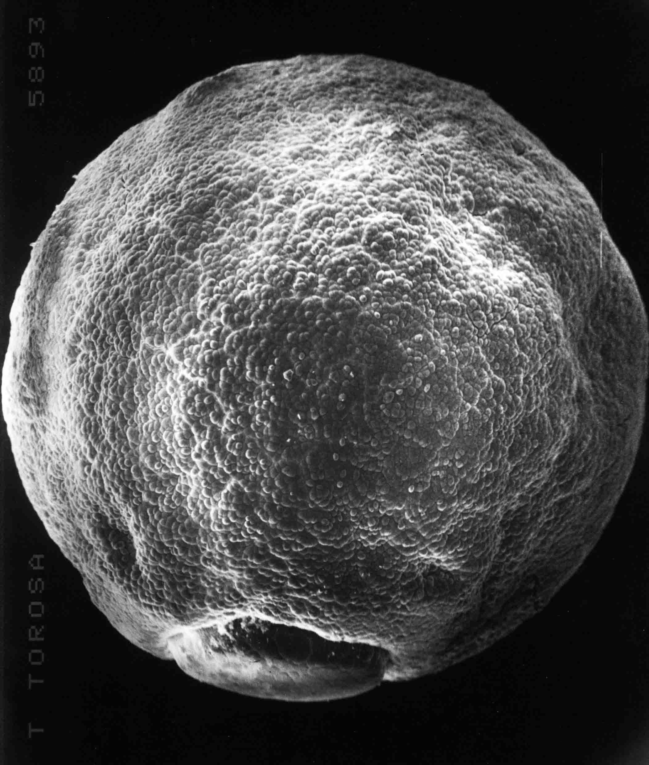 Dorsal surface view of a stage 12 embryo (49X). The blastopore is at the posterior end (bottom).
The dorsal surface is prospective neural plate.
Dorsal surface view of a stage 12 embryo (49X). The blastopore is at the posterior end (bottom).
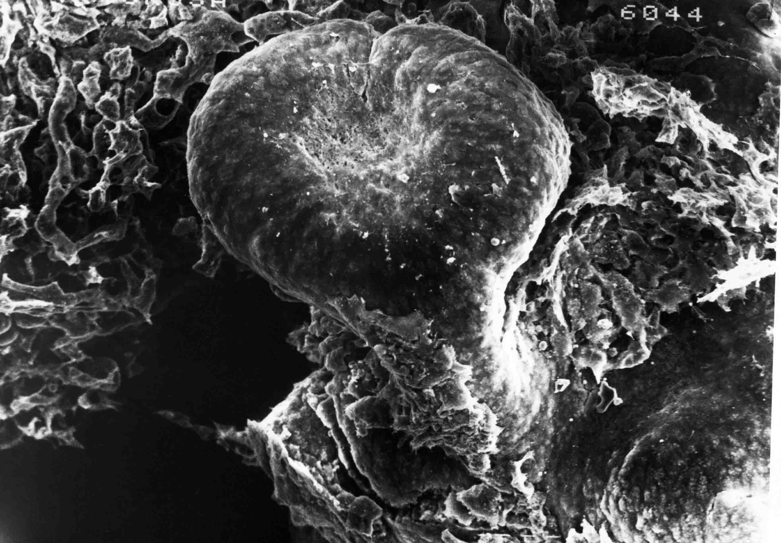
The dorsal surface is prospective neural plate.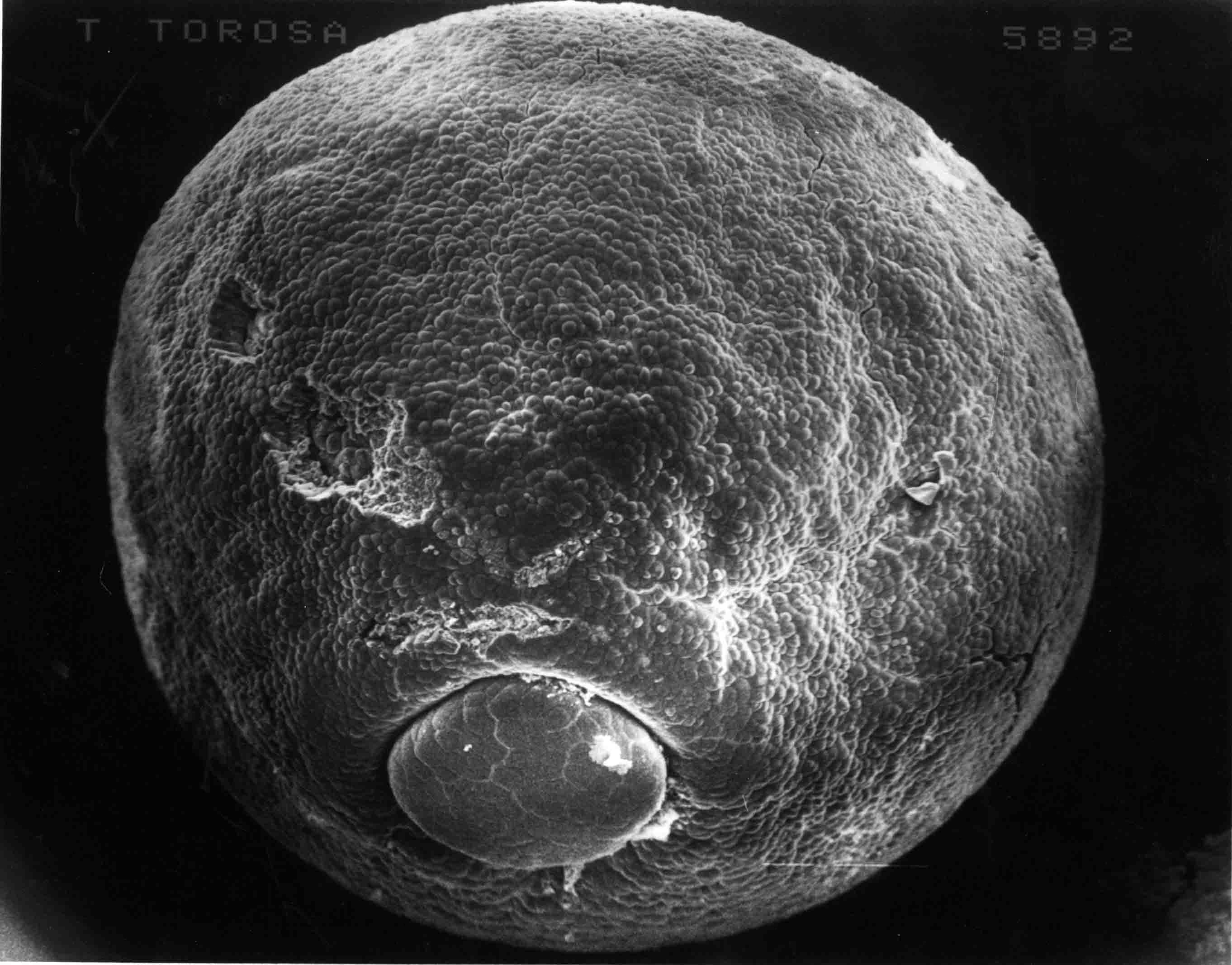 Dorsal, slightly posterior surface view of a stage 13 embryo. The yolk plug is smaller than at
stage 12. The endoderm cells of the yolk plug continue to involute until they are all inside.
The forming neural plate extends posterior and a bit ventral to the yolk plug.
Dorsal, slightly posterior surface view of a stage 13 embryo. The yolk plug is smaller than at
stage 12. The endoderm cells of the yolk plug continue to involute until they are all inside.
The forming neural plate extends posterior and a bit ventral to the yolk plug.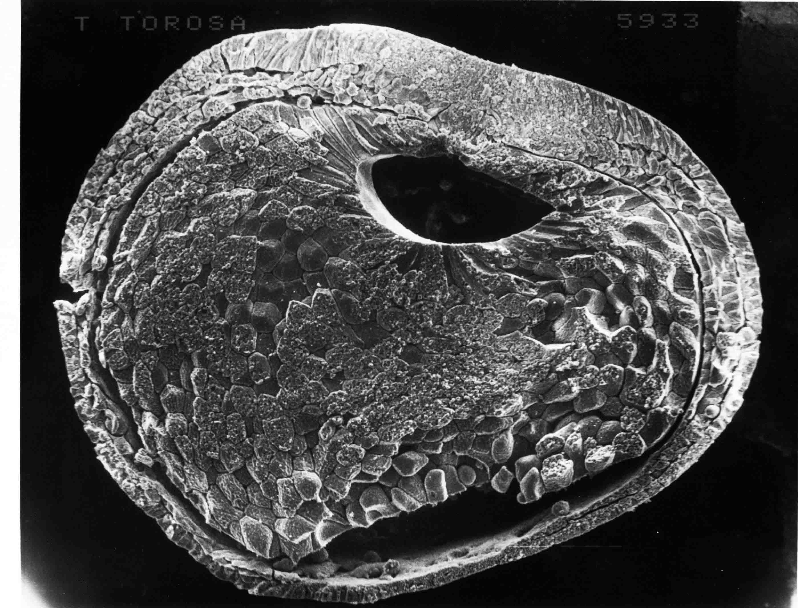 This is a cross section of a stage 13+ embryo (58X). At the midline above the endoderm of the gut
cavity is the notochord, and above that is the notoplate which is the midline part of the neural
plate. Notoplate and notochord are tightly fused. A bilaminar somitomere lies to each side of the
notochord. The somitomeres and the neural plate above them extend nearly to the equator of the embryo
at this stage.
This is a cross section of a stage 13+ embryo (58X). At the midline above the endoderm of the gut
cavity is the notochord, and above that is the notoplate which is the midline part of the neural
plate. Notoplate and notochord are tightly fused. A bilaminar somitomere lies to each side of the
notochord. The somitomeres and the neural plate above them extend nearly to the equator of the embryo
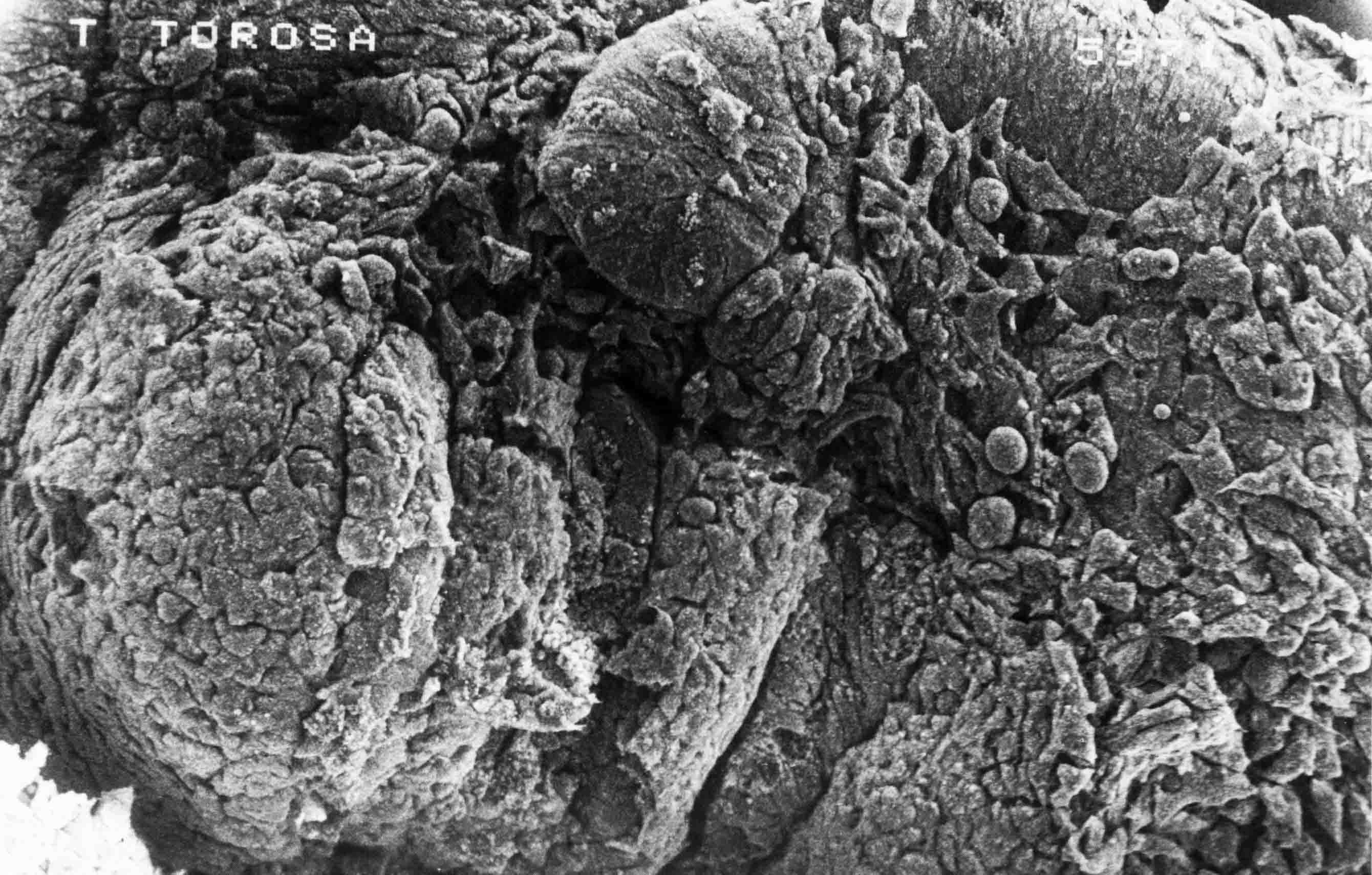
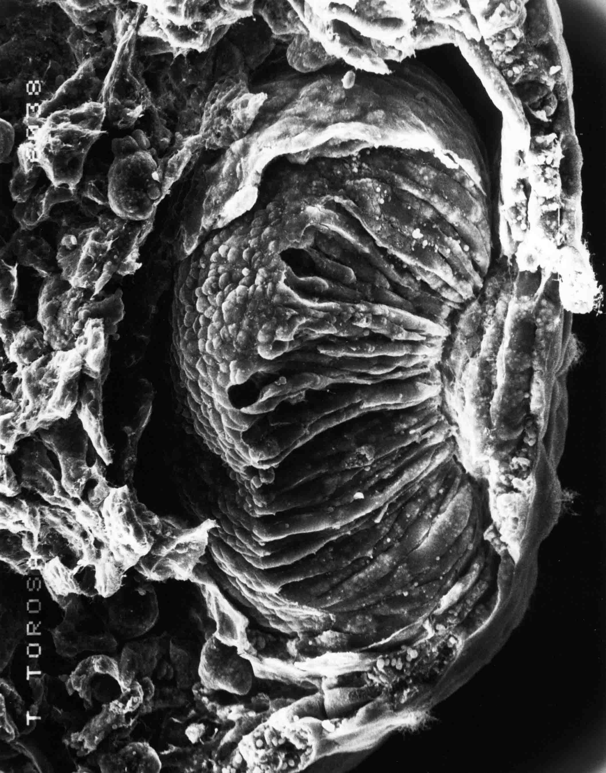
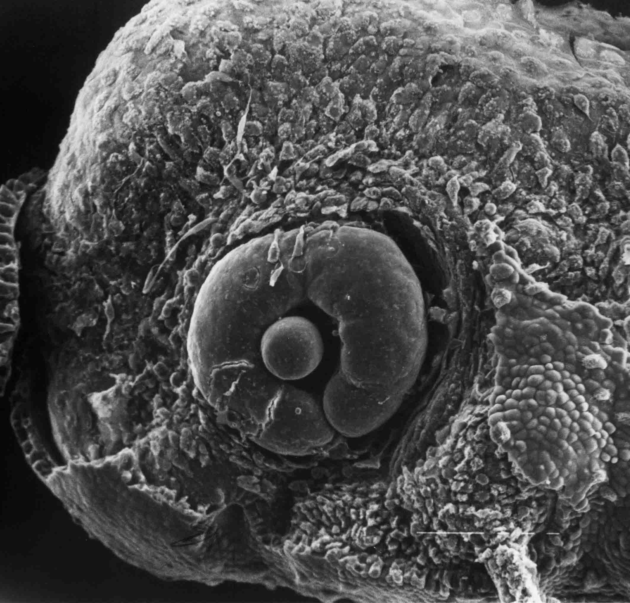
at this stage.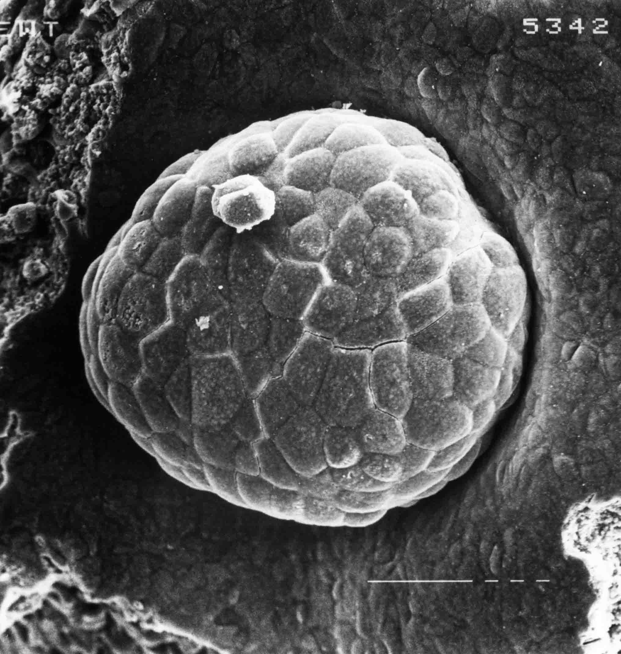 Stage 13; 149X. The yolk plug of a stag 13 embryo. After stage 12, the yolk plug gets
progressively smaller. The endodermal cells of the yolk plug continue involuting beneath the
blastopore lips. The circular lip gets smaller as the cells that compose the lip continue to
involute. Convergent-extension helps close the blastopore as well.
Stage 13; 149X. The yolk plug of a stag 13 embryo. After stage 12, the yolk plug gets
progressively smaller. The endodermal cells of the yolk plug continue involuting beneath the
blastopore lips. The circular lip gets smaller as the cells that compose the lip continue to
involute. Convergent-extension helps close the blastopore as well.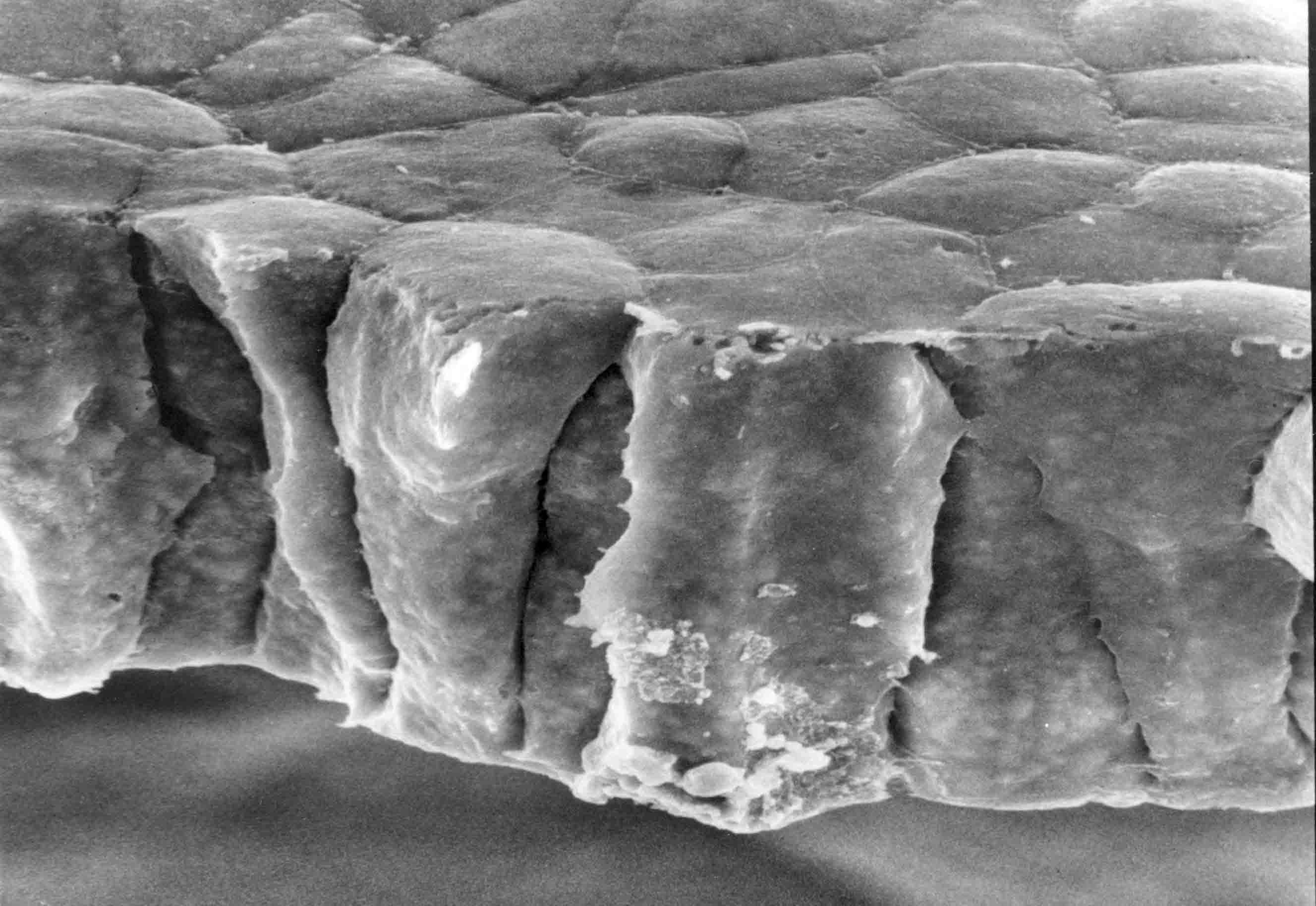 Stage 13; 700X. The cut edge of the neural plate at stage 13. Apical ends of the neural plate cells
are upward; basal ends of these epithelia cells are downward. The shapes of these cells strongly
suggest that contraction of the apical ends of these cells is not the leading force in the
elongation of the cells that is occurring. There are many cell processes from the sides and base of
the cells.
Stage 13; 700X. The cut edge of the neural plate at stage 13. Apical ends of the neural plate cells
are upward; basal ends of these epithelia cells are downward. The shapes of these cells strongly
suggest that contraction of the apical ends of these cells is not the leading force in the
elongation of the cells that is occurring. There are many cell processes from the sides and base of
the cells. 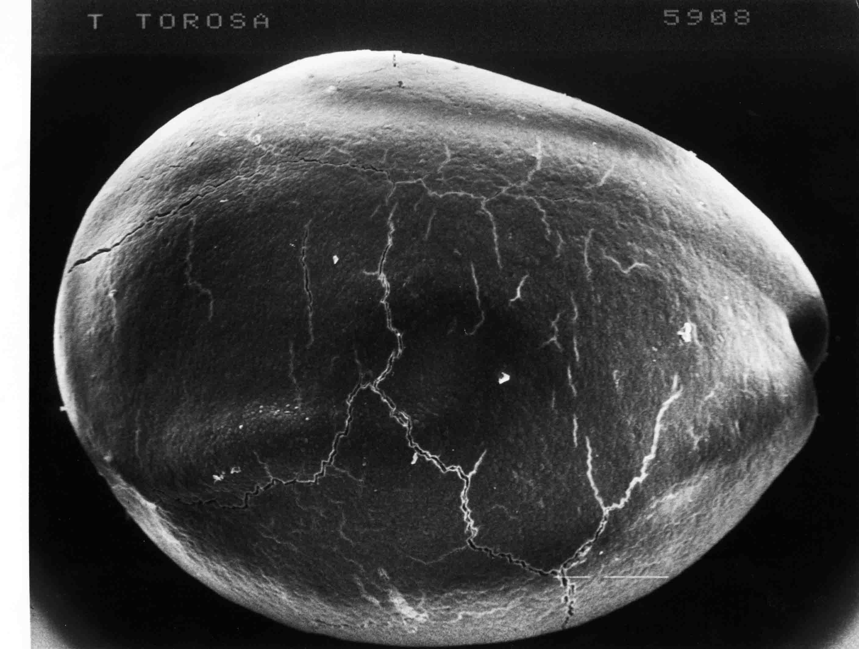 Stage 14, 43X, surface view of neural plate. Posterior is to the right. By stage 14, the neural
plate is a nearly flat disc the diameter of the embryo.
Stage 14, 43X, surface view of neural plate. Posterior is to the right. By stage 14, the neural
plate is a nearly flat disc the diameter of the embryo. 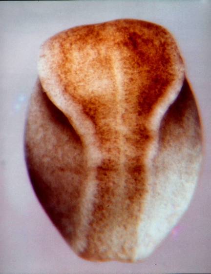 This is a color photograph of a stage 15 embryo in dorsal view. A color photograph rather than an SEM
shows best the natural variegation in pigmentation among the cells of the neural plate. The neural
folds have started to rise around the neural plate and the plate has begun to elongate. The future
brain is the wider part of the plate. The longer, narrower part of the plate is the spinal cord area.
Along the midline, a less pigmented streak runs from the posterior end of the embryo (bottom) to the
anterior tip of the future midbrain in the brain plate. This streak is the notoplate, a part of the
neural plate that lies directly above the notochord in the underlying mesoderm. The notoplate assists
in plate elongation.
This is a color photograph of a stage 15 embryo in dorsal view. A color photograph rather than an SEM
shows best the natural variegation in pigmentation among the cells of the neural plate. The neural
folds have started to rise around the neural plate and the plate has begun to elongate. The future
brain is the wider part of the plate. The longer, narrower part of the plate is the spinal cord area.
Along the midline, a less pigmented streak runs from the posterior end of the embryo (bottom) to the
anterior tip of the future midbrain in the brain plate. This streak is the notoplate, a part of the
neural plate that lies directly above the notochord in the underlying mesoderm. The notoplate assists
in plate elongation.
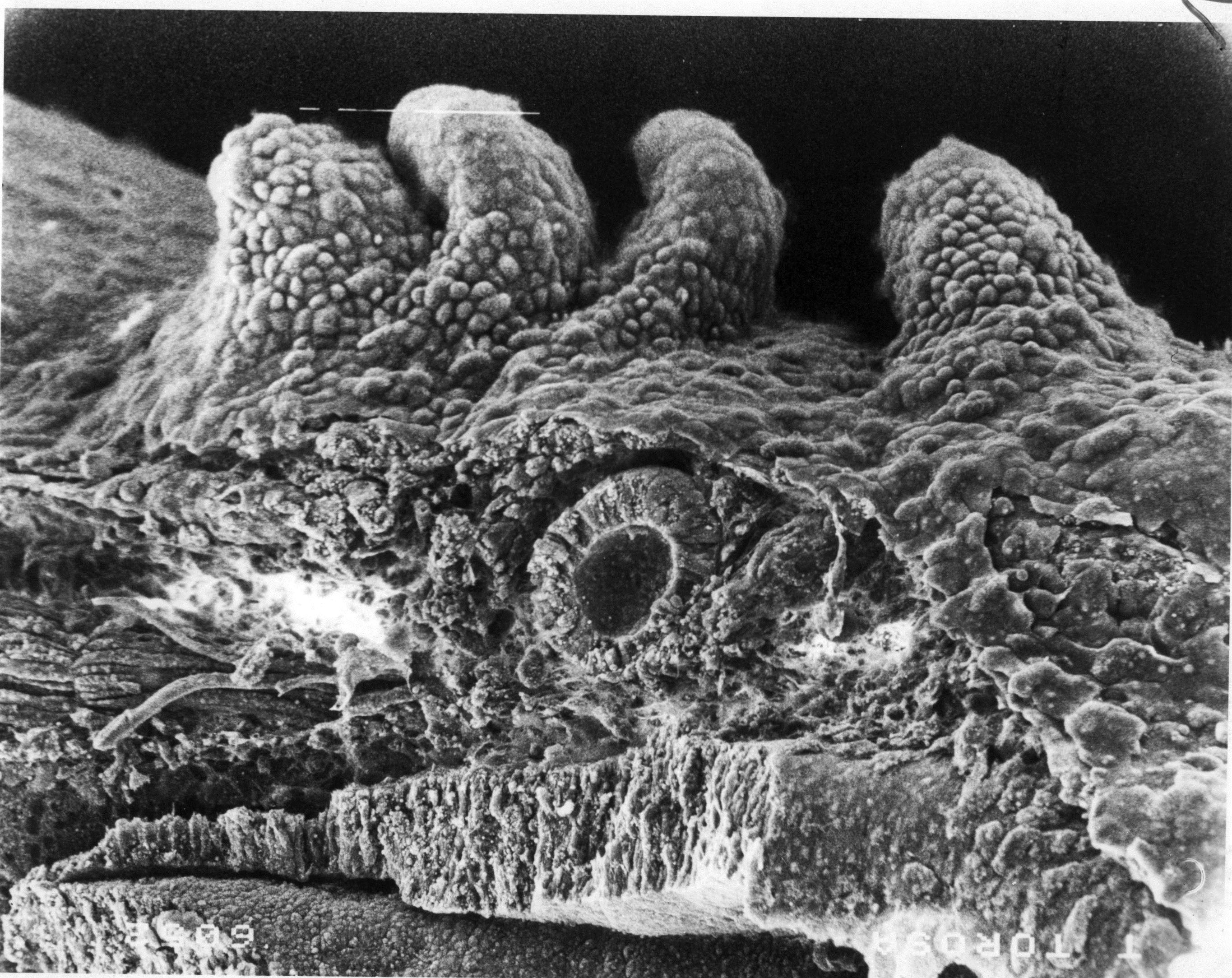
Neurulation from stages 12 to 20 is discussed for this newt on this web site at Neurulation. Somitomeres, the segmental precursors of somites, are apparent in the newt embryo during neurulation stages. Somitomere formation is discussed in the newt and in other vertebrates at Somitomeres, Somites. The collection of newt sem's number two or three hundred. There are many cases with consistent results, making the analysis of somitomeres in this species very robust. A couple of examples follow. 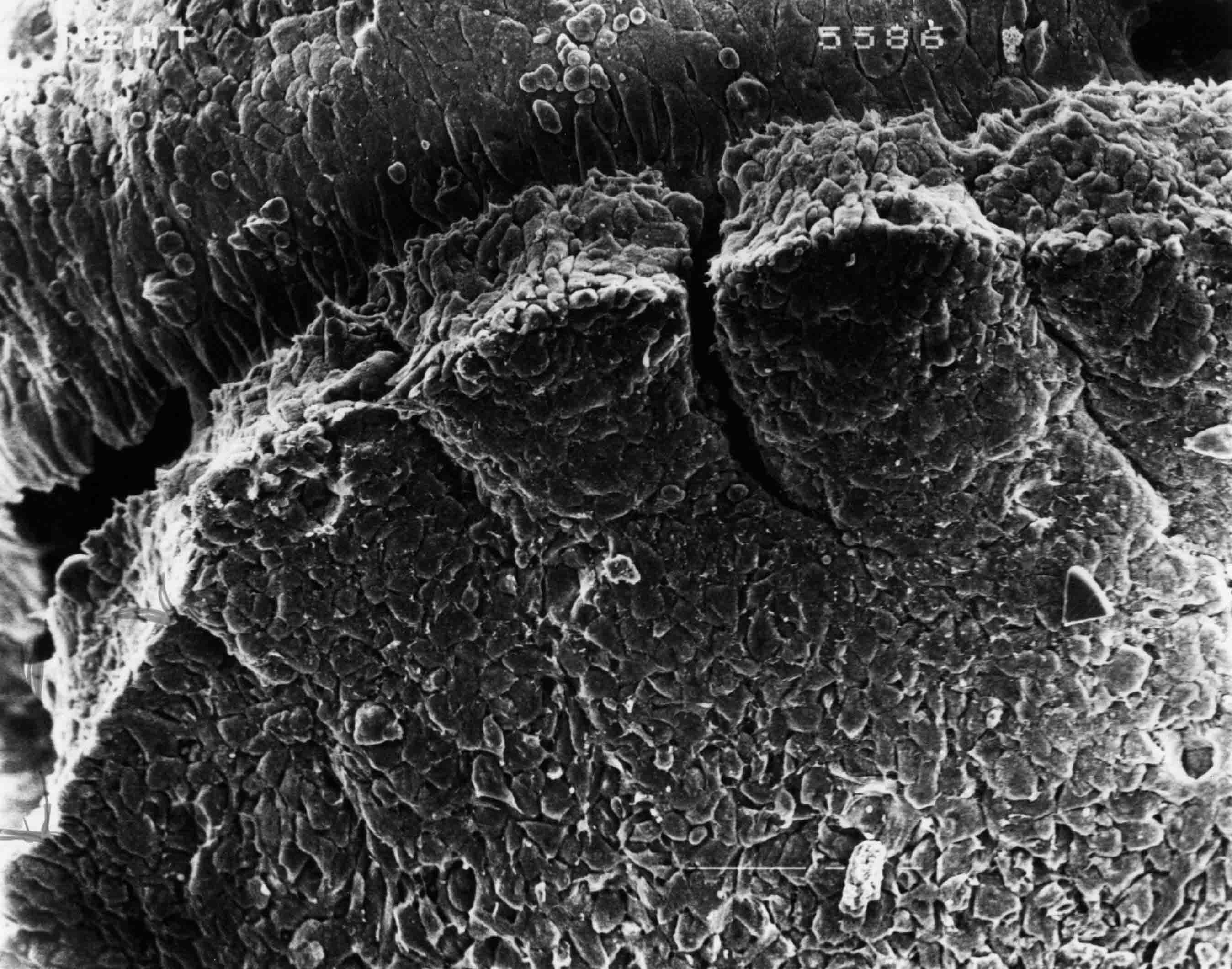 Recently formed somites in a stage 18 newt embryo (111X).
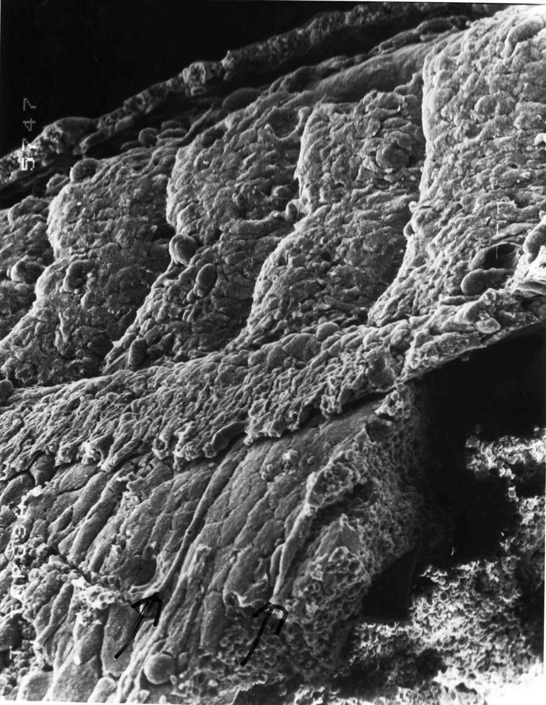
 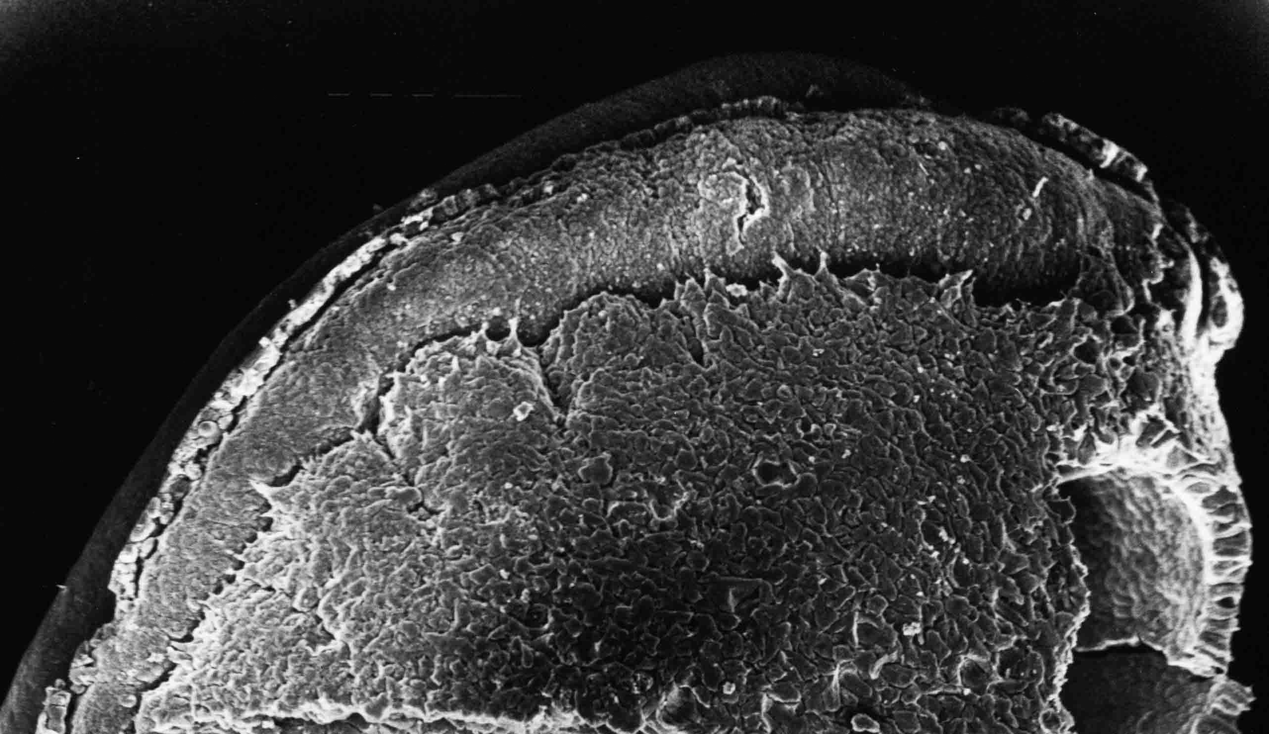 Lateral view of a stage 19 embryo with epidermis removed (57X)
A row of somites with somitomeres at each end.
Lateral view of a stage 19 embryo with epidermis removed (57X)
A row of somites with somitomeres at each end. 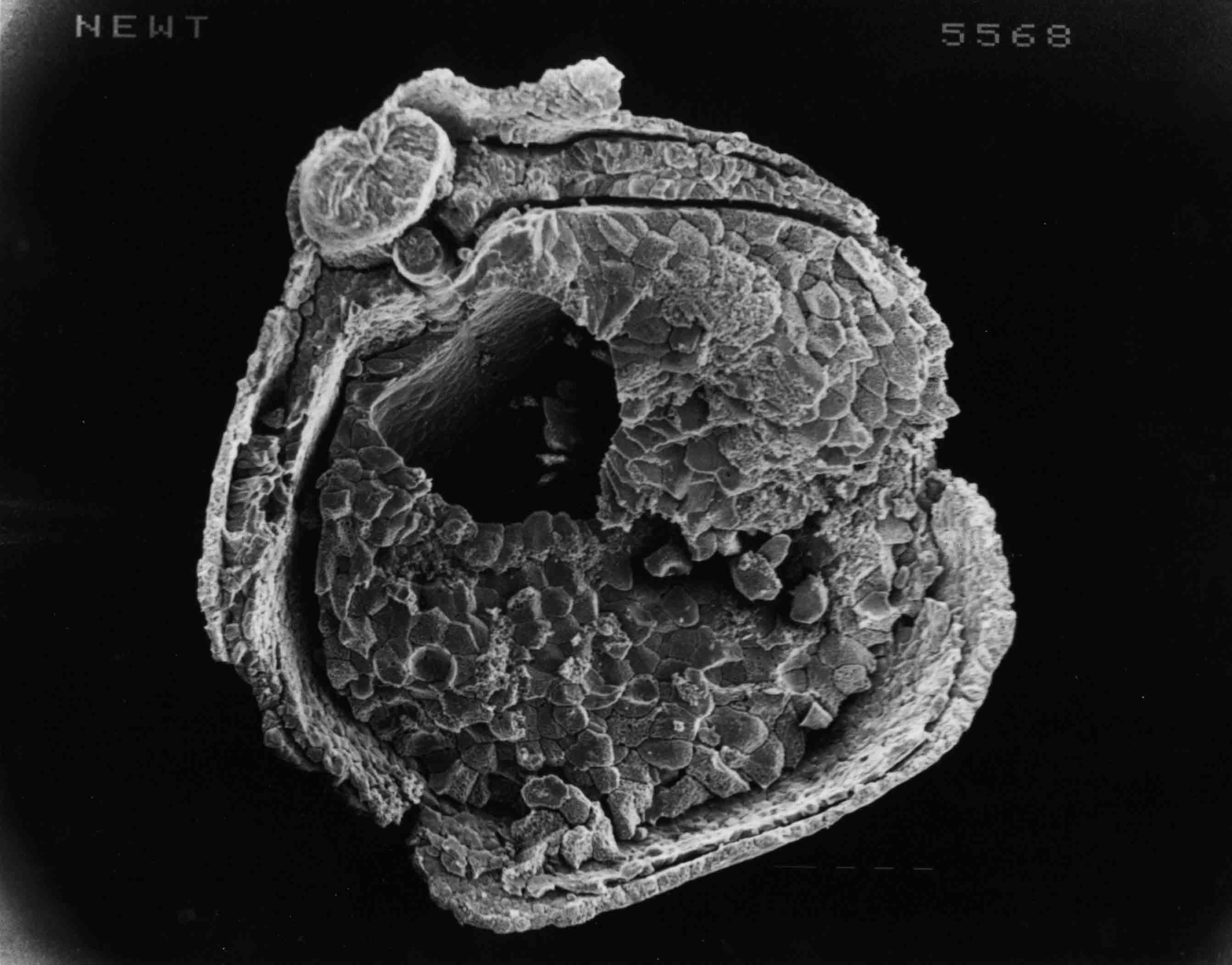 A cross section through a stage 18 embryo (38X). To the right of the neural tube is a somite. The
mesoderm layer consists of the notochord at the midline beneath the neural tube, then the somite
which is adjacent to intermediate mesoderm then lateral plate mesoderm; somatic lateral plate
beneath the epidermis and splanchnic lateral plate against the endoderm.
A cross section through a stage 18 embryo (38X). To the right of the neural tube is a somite. The
mesoderm layer consists of the notochord at the midline beneath the neural tube, then the somite
which is adjacent to intermediate mesoderm then lateral plate mesoderm; somatic lateral plate
beneath the epidermis and splanchnic lateral plate against the endoderm. 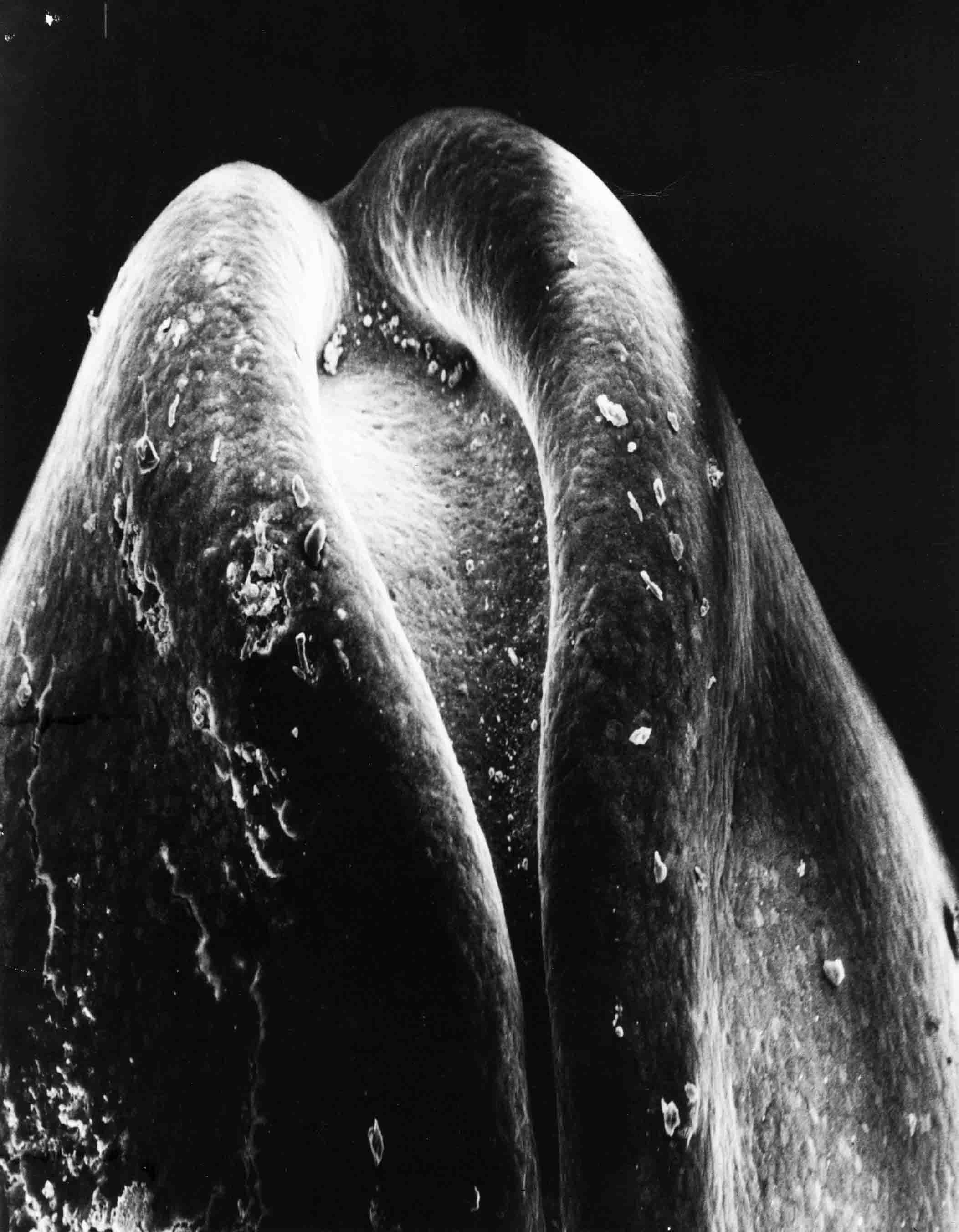 The closing brain plate of a stage 19 newt embryo (75X).
 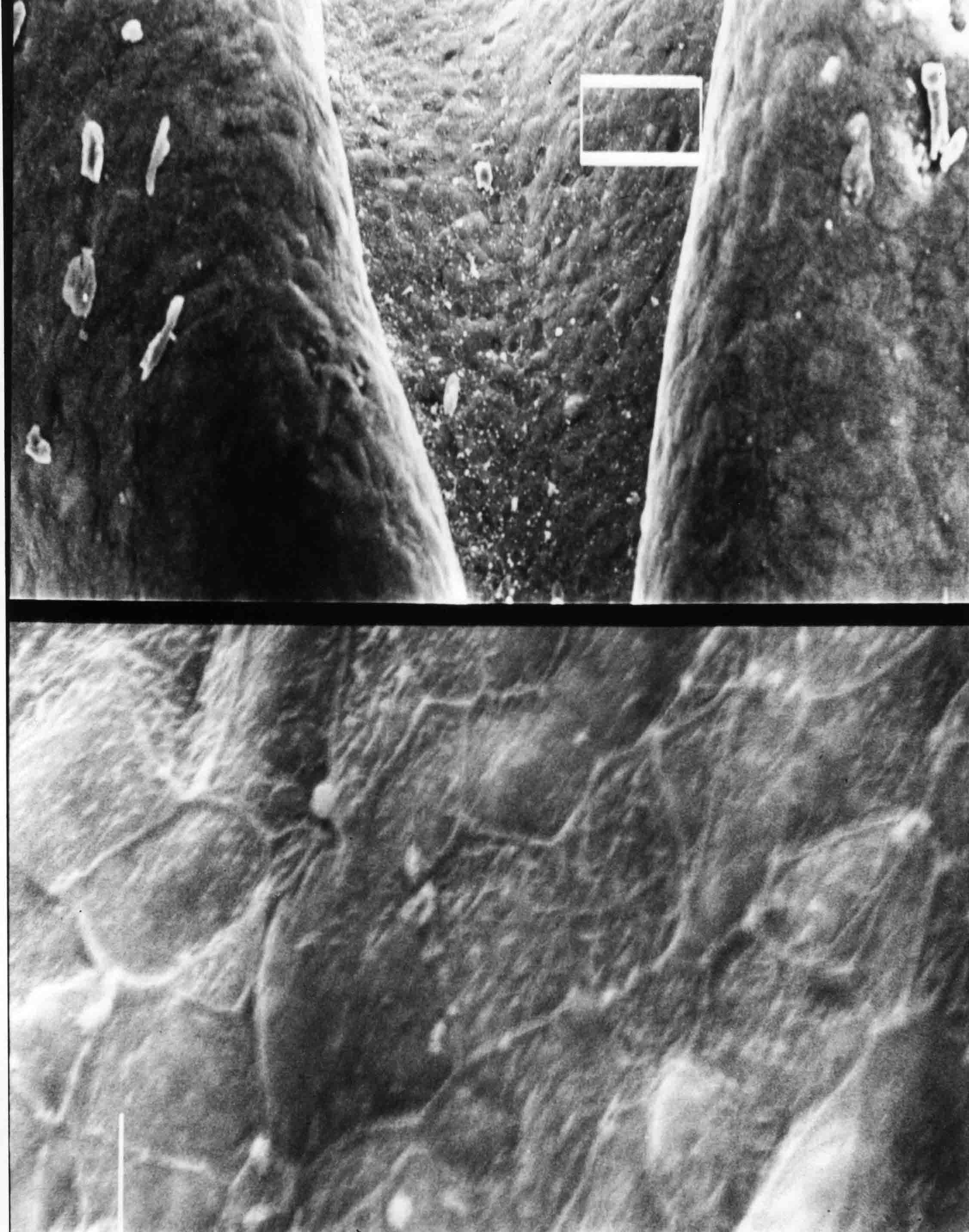 In the upper part of this sem, the box encloses a group of brain plate cells (150X). This group of
cells is shown at 1500X in the bottom part of the sem. Size and orientation of the apical ends of
these cells varies. (Stage 19).
In the upper part of this sem, the box encloses a group of brain plate cells (150X). This group of
cells is shown at 1500X in the bottom part of the sem. Size and orientation of the apical ends of
these cells varies. (Stage 19).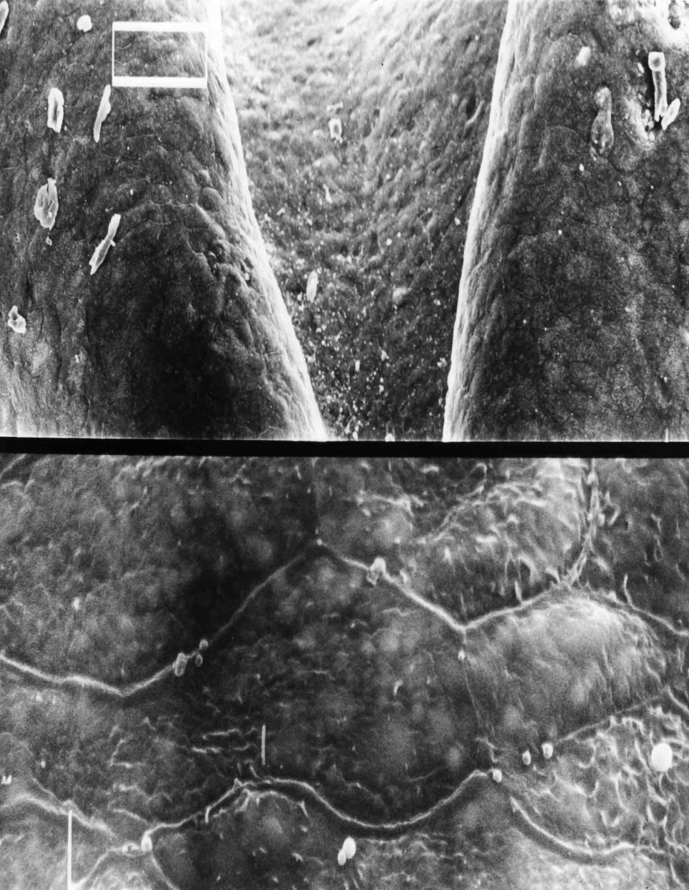 The boxed area in the upper photo encloses neural fold cells (stage 19) (150X). Apical serfaces of
these cells are shown in the lower part of the sem at 1500X.
The boxed area in the upper photo encloses neural fold cells (stage 19) (150X). Apical serfaces of
these cells are shown in the lower part of the sem at 1500X.  By stage 20, the neural folds have come in contact the length of the nervous system as shown in this
example (42X). The anterior end i at left. After fixation, the epidermis on the left side was
removed, revealing to the side of the neural tube somites and somitomeres. Even though the folds
are in contact, they have not yet fused, though they do by stage 21.
By stage 20, the neural folds have come in contact the length of the nervous system as shown in this
example (42X). The anterior end i at left. After fixation, the epidermis on the left side was
removed, revealing to the side of the neural tube somites and somitomeres. Even though the folds
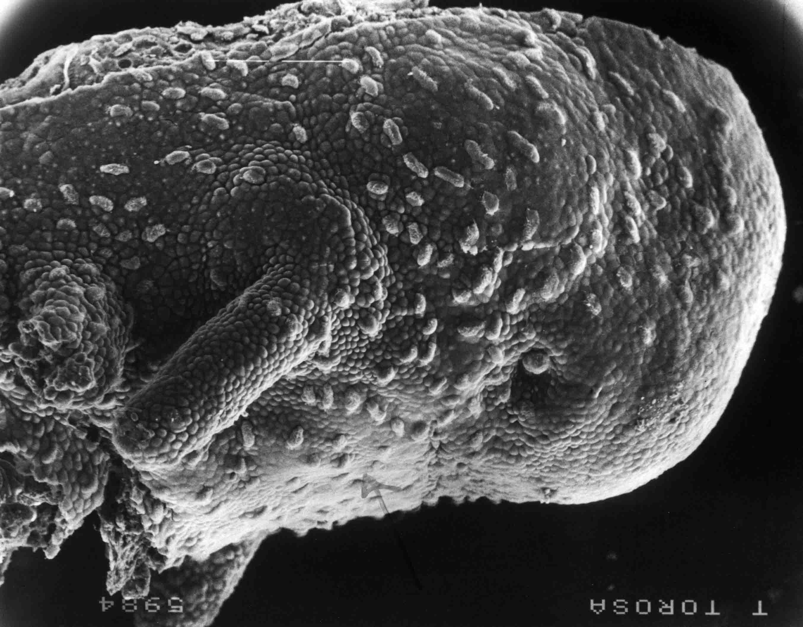
are in contact, they have not yet fused, though they do by stage 21. The cranial segments of the newt are readily apparent. The illustration below (from 1984 A.G. Jacobson and S. Meier. Dev. Biol. 106:181-193. link .) is of a stage 23 embryo, which is the oldest stage at which the neural crest has not yet migrated over and obscured the somitomeres. The left side of the embryo is viewed in a scanning electron micrograph (X58) The epidermis has been removed. 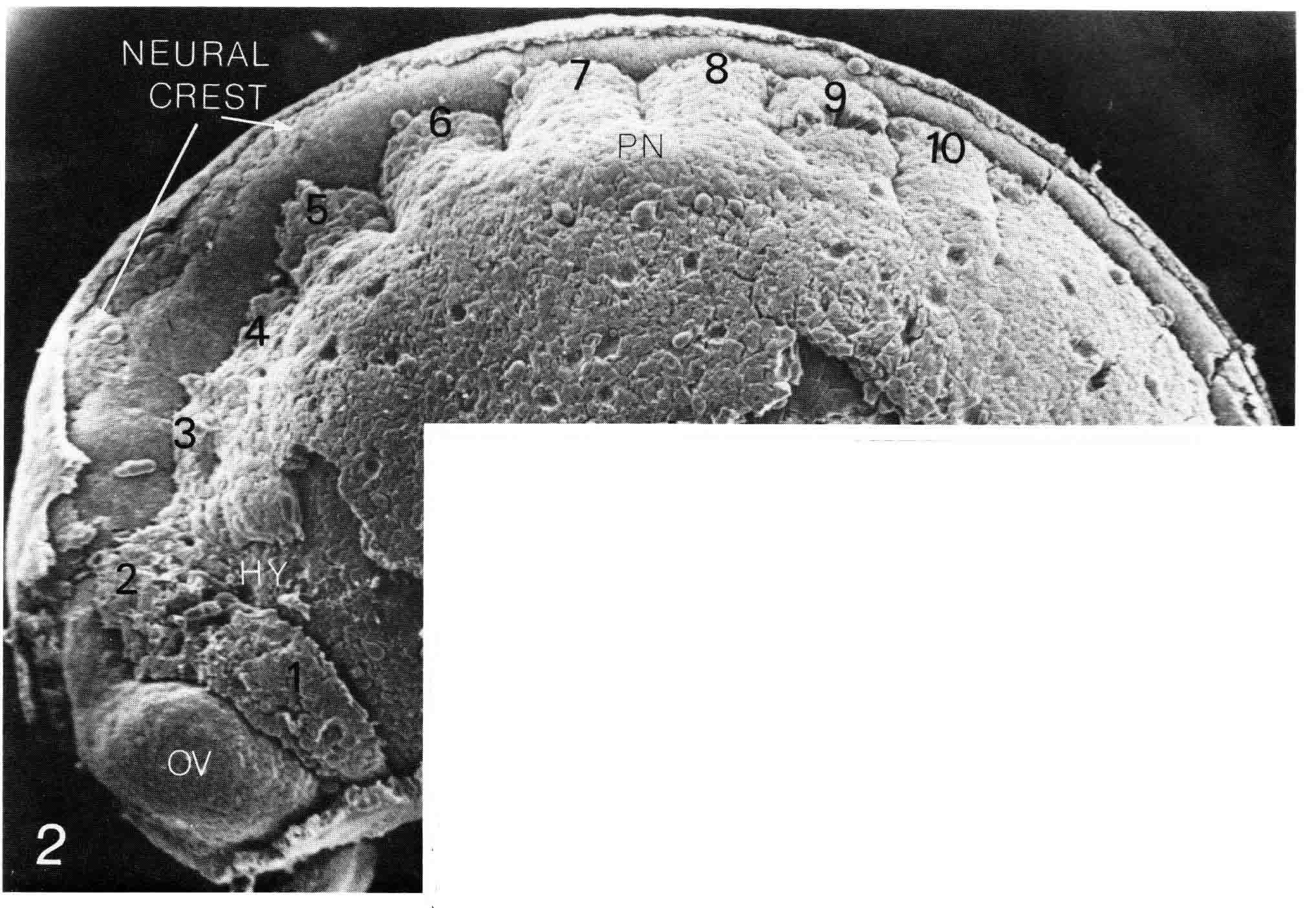
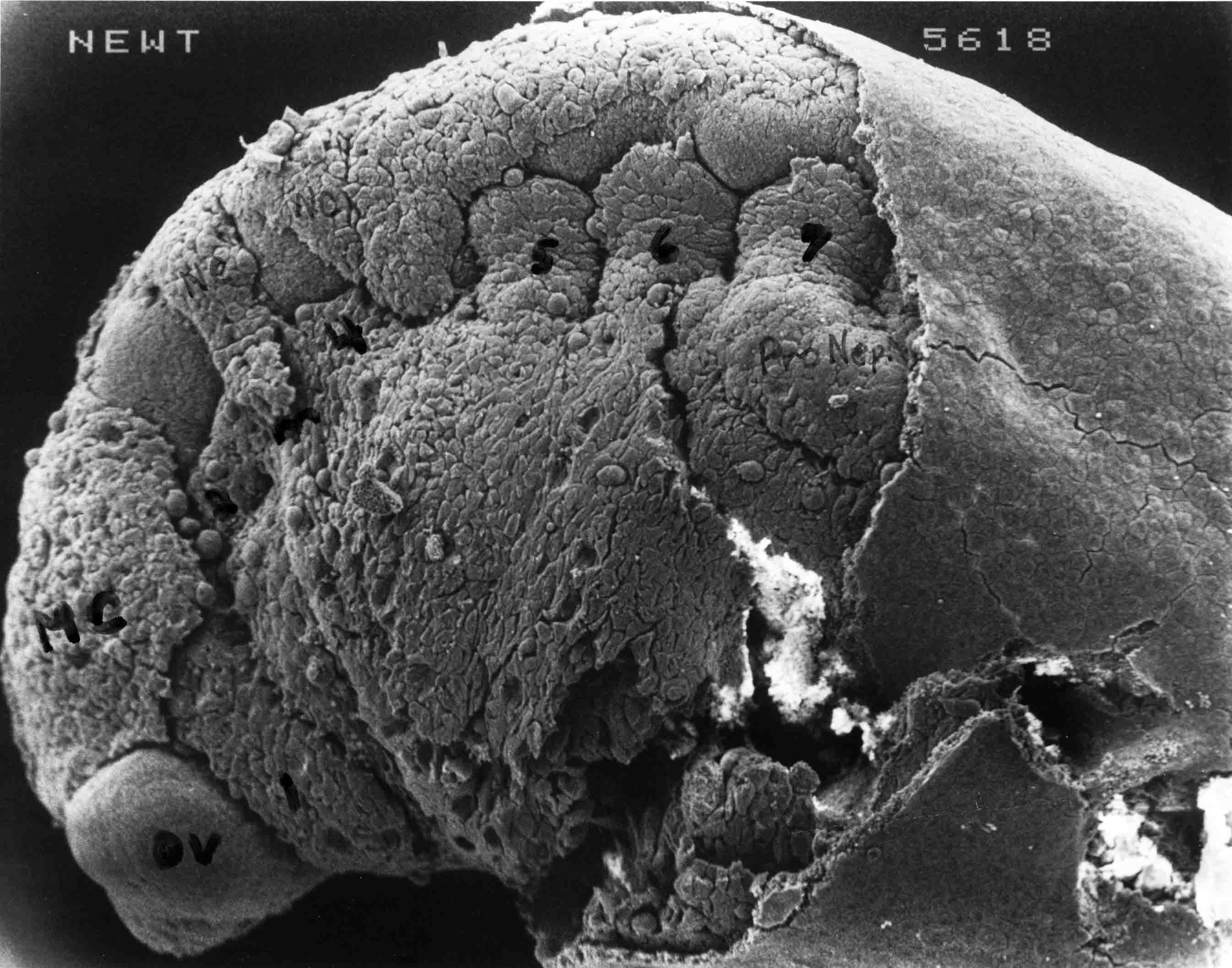
At stage 24 (below), the neural crest migrates from the dorsal midline. The mesencephalic crest is first to migrate and by stage 24 has covered much of the forebrain. 
|