
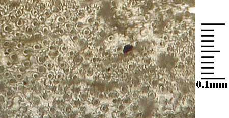
The distinctions in Sporobolus lemma vestiture observed with SEM scans can also be observed at the lower magnifications of a compound microscope, at least at 400X magnification. Although this is not the best method of viewing the micromorphology of the lemma surface, once the primary distinctions have been determined with SEM scans (shown immediately below), it can be used to approximate matches in the absence of access to an SEM.
 |  |  |
|---|---|---|
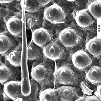
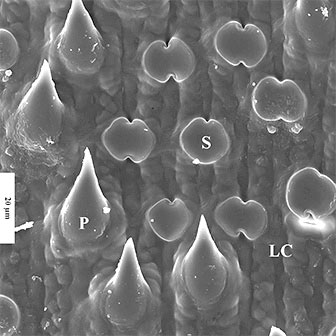
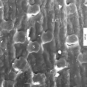
| S. vaginiflorus with hooks | S. clandestinus prickles & silica cells | S. compositus var. compositus silica cells |
| S = silica cell, LC = long cell [scale bar is 20µm] | ||
To determine the feasibility of this approach lemmas were selected from 3 major types:
The lemmas, which were tightly folded along the midrib, were cut into small flat segments and then softened in a water/aerosol solution. These were then placed with the abaxial surface up on a glass slide and covered with a coverslip. Unlike views with a dissecting scope with reflected light or coated with platinum with the SEM, light with the compound microscope must pass through the lemma. Fortunately this did pass sufficient light to allow focusing on the abaxial upper surface details. With S. clandestinus and S. compositus var. macer it also permitted greater detail of the strongly sinuate sidewalls of the long cells than was obtained with the earlier imaged coated SEM cells.
Although many details were not not clear at 100X, overall surface distinctions were apparent (given that the basic structure is already known). The images below at the same scale show (a) with S. vaginiflorus the dense covering of hooks (dark round dots within a larger circular base), the long cells only barely visible; (b) the strongly sinuate long cells of the other two taxa; (c) pale, short and somewhat rounded silica cells regularly spaced among the long cells prominent with S. compositus and (d) the abundant larger slighly oval dark cells on the surface of S. clandestinus (i.e., prickles, with the translucent prickle itself not visible at this magnification).
 |  |
|---|---|
| S. vaginiflorus | S. clandestinus |
 |
|
| S. compositus var. macer | |
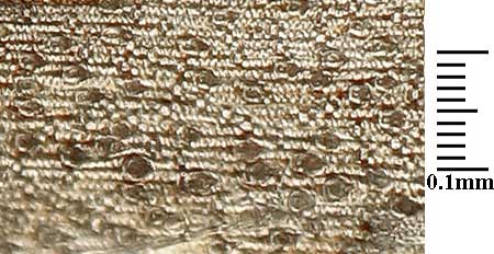
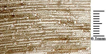
The primary cell distinctions could be seen at 400X. The images below all reflect 0.14 mm high lemma sections at the same scale. Depth of field was critical at 400X, but also permitted viewing the cells at different focal lengths, especially important for hook and prickles. [Click on an image for a larger view.]

The same area of S. vaginiflorus at two focal lengths. The double-headed arrows are at the same place in the two images. The red circles show the acute top ‘hook’ of the cell more or less in focus in the same area as the pink circles (unfocused) on the adjacent image. A macrohair is in focus on the right image.

Prickles of S. clandestinus in the same area as that shown in the image below. Focus here is on the translucent prickle tops. Note the dark basal portion of the cell (which for whatever reason does not transmit light from below).

S. clandestinus prickle bases (P) at the same focal plane as the pale/whitish silica cells (S) and the long cells (L). The translucent prickle tops are not visible against the focused dark background.
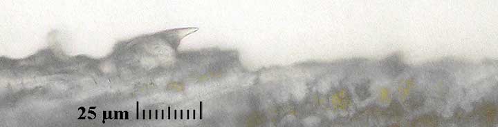
Lateral view of S. clandestinus prickle.

All features of S. compositus can be seen on a single focal plane. The sinuate pattern of the long cells (L) is slightly different from that of S. clandestinus, more open, and the silica cells (S) are larger and seem to occur regularly between the long cells.