
Sample Images
Provided below are links to some of the outstanding images collected using the Center's microscopes:
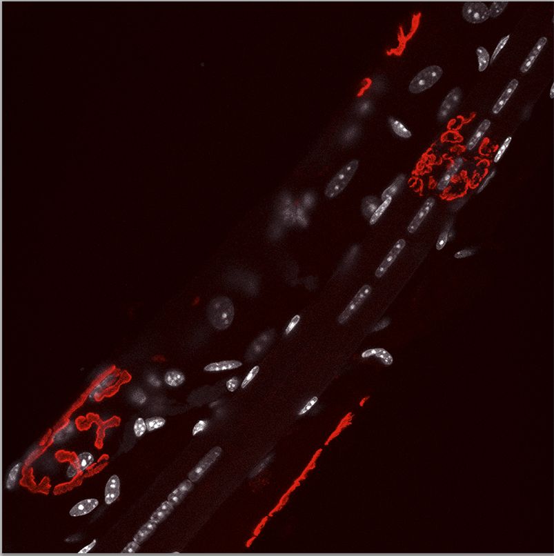 |
| Sample 1. Muscle fiber under repair following damage. Maximum projection of image stack. Fiber run along an angle of approximately 45 degrees. Acetylcholine receptors (red in image) have been labeled with rhodamine bungarotoxin. Nuclei (shown as grey) are labeled with DAPI. A string of "central nuclei" are located in one of the fibers. Other muscle nuclei are subsacrolemmal. Image courtesy of Matt Lee. |
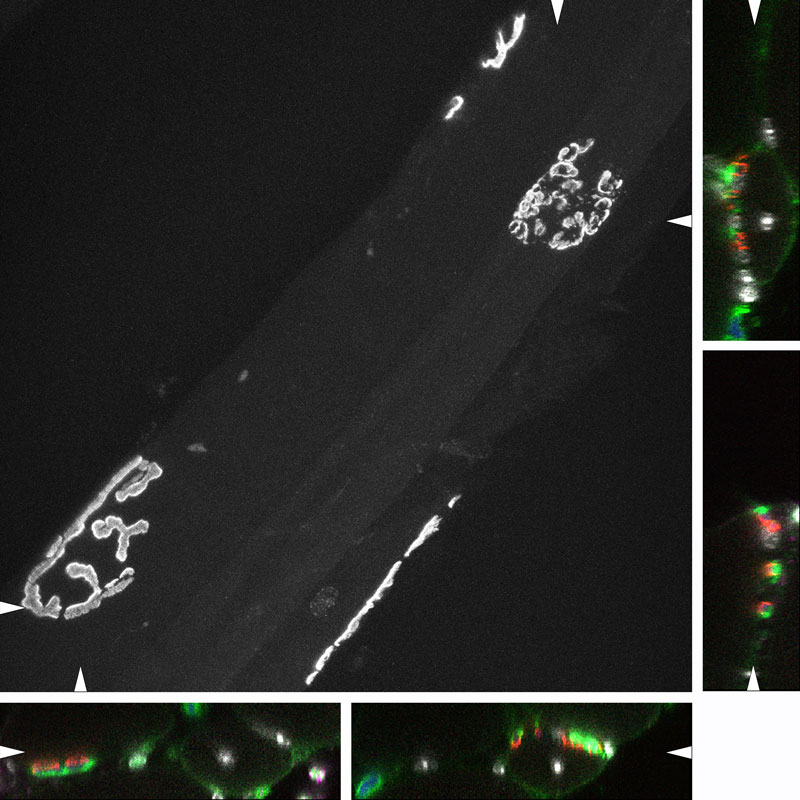 |
| Sample 2. Images of the same field as in Sample 1. The major image is a maximum projection of only the acetylcholine receptor labeling. Shown to the sides are portions of x-z and y-z projections at the levels indicated by the arrowheads. These projections clearly show the central location of the nuclei. Images courtesy of Matt Lee. |
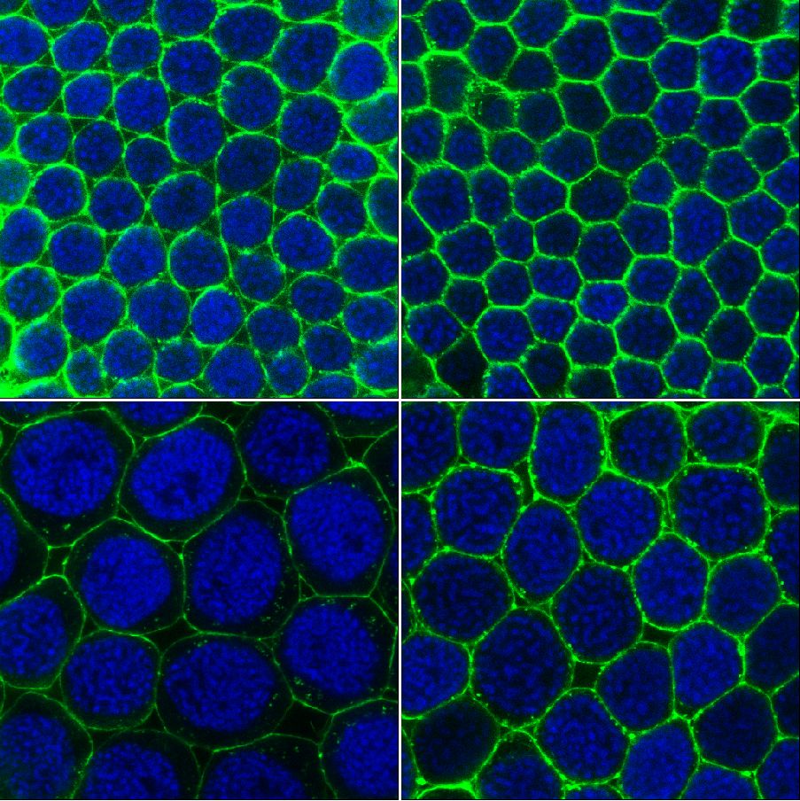 |
| Sample 3. Confocal images of developing grasshopper oocytes. Alexa 488 phalloidin (green in image) outlines individual follicle cells and DAPI (blue) labels the nuclei of follicle cells within each oocyte. Treatment of grasshopper with juvenile hormone allows one to examine the influence of this hormone on patency, i.e. formation of gap spaces between follicle cells to permit the uptake of yolk proteins into the oocyte. Images of Tina Taub-Montemayor. |
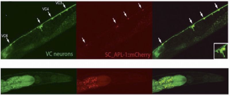 |
| Sample 4. Ventral cholinergic neurons (VC neurons) in C. elegans marked with transgenic GFP (left panel) also express mCherry tagged APP driven from a neuronal promoter (middle panel). Far right panel merges the other two images. The inset clearly shows the colocalization in VC neuron 5. In this figure, the neurons are still alive. The lower panels are head shots of the worm. The nose is pointing to the right. APP is selectively accumulating in the VC neurons and not elsewhere in the body. Image of Ashley Crisp. |
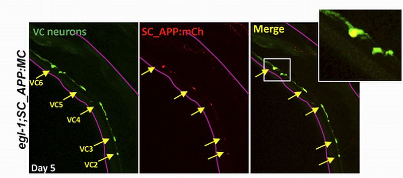 |
| Sample 5. Ventral cholinergic neurons (VC neurons) in C. elegans expressing mCherry-tagged APP. Six cholinergic neurons in the ventral nerve chord of the worm (outlined in magenta) are labeled with transgenic GFP. Even though the APP gene is being overexpressed throughout the nervous system, it appears to selectively accumulate in VC neurons, as seen by mCherry fluorescence. Asterisks indicate autoflourescence in the gut. Since this worm was of the egl-(lf) background, VC4&5, which normally degenerate when overexpressing APP are spared from degeneration. This shows that APP-mediated death of these VC neurons is upstream of this cell death gene. These neurons still accumulate APP protein over time (inset) even without egl-1. Scale bar 40 µm. Image of Ashley Crisp. |
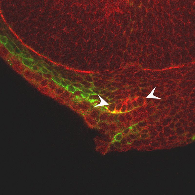 |
| Sample 6. Optical sections of fluorescence in situ hybridization verifies the expression expression pattern of pax9, a transcription factor at 40 hours postfertilization (hpf) in a zebrafish head. Lateral view of the zebrafish head; mRNA localization detected by fluorescence of the NBT/BCIP precipitate (red) and anti-EGFP immunostaining labels cranial neural crest cells (CNCC) in the fli1:EGFP transgenic background (green). Arrowheads indicate CNCC expression domains. Image of Mary Swartz. |
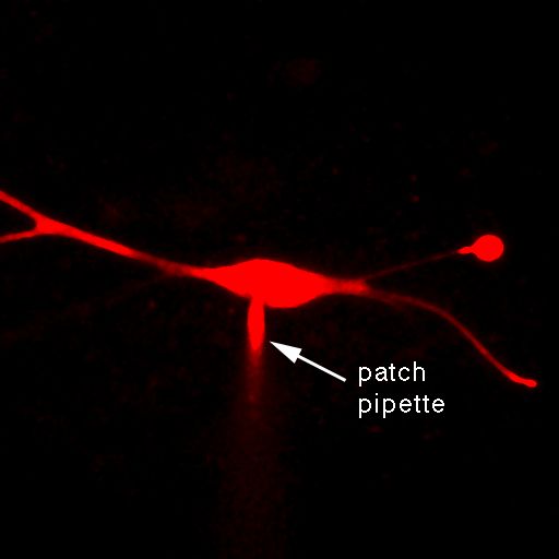 |
| Sample 7. A single. Living neuron in the medial superior olive (MSO) of brain slice from the gerbil. Neuron has been filled with the dye Alexa 594 through a patch clamp pipette visible in the image. Maximal projections of an image stack. Image from Kwang Woo Ko. |